Vascularisation Strategies in Stem Cell-Based Bone Tissue Engineering
DOI:
https://doi.org/10.5281/zenodo.22274391Keywords:
Bone tissue engineering; Vascularisation; Mesenchymal stromal cells; Angiogenesis; Scaffold architectureAbstract
Background: Constructs larger than the diffusion limit die at their centre, so vascularisation rather than osteogenic
induction has been the rate-limiting step in stem cell-based bone tissue engineering.
Objective: To assess the evidence behind vessel-instructive biology, scaffold architecture, growth-factor dose, co-culture
prevascularisation, printed channels and surgically axialised vascular beds.
Methods: Narrative review of rodent, rabbit and sheep implantation studies, in vitro co-culture and printing work, human
biopsy series and clinical case reports.
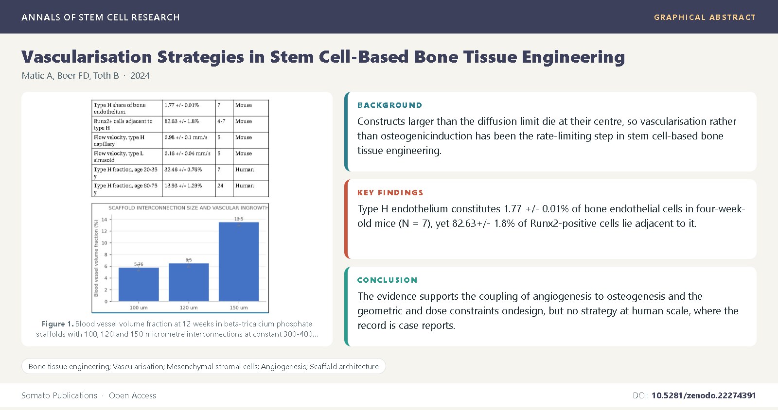
Findings: Type H endothelium constitutes 1.77 +/- 0.01% of bone endothelial cells in four-week-old mice (N = 7), yet 82.63
+/- 1.8% of Runx2-positive cells lie adjacent to it. In rabbit femoral condyles, vessel volume fraction at twelve weeks was 13.5
+/- 0.58% with 150 micrometre interconnections against 5.76 +/- 0.52% at 100 micrometres (N = 6).
Conclusions: The evidence supports the coupling of angiogenesis to osteogenesis and the geometric and dose constraints on
design, but no strategy at human scale, where the record is case reports.
References
Simunovic, F., Finkenzeller, G. (2021) Vascularization strategies in bone tissue engineering. Cells, 10(7): 1749.
Rouwkema, J., Rivron, N.C., van Blitterswijk, C.A. (2008) Vascularization in tissue engineering. Trends in Biotechnology, 26(8): 434-441.
Rouwkema, J., Khademhosseini, A. (2016) Vascularization and angiogenesis in tissue engineering: beyond creating static networks. Trends in Biotechnology, 34(9): 733-745.
Filipowska, J., Tomaszewski, K.A., Niedzwiedzki, L., Walocha, J.A., Niedzwiedzki, T. (2017) The role of vasculature in bone development, regeneration and proper systemic functioning. Angiogenesis, 20(3): 291-302.
Grellier, M., Bordenave, L., Amedee, J. (2009) Cell-to-cell communication between osteogenic and endothelial lineages: implications for tissue engineering. Trends in Biotechnology, 27(10): 562-571.
Kusumbe, A.P., Ramasamy, S.K., Adams, R.H. (2014) Coupling of angiogenesis and osteogenesis by a specific vessel subtype in bone. Nature, 507(7492): 323-328.
Ramasamy, S.K., Kusumbe, A.P., Wang, L., Adams, R.H. (2014) Endothelial Notch activity promotes angiogenesis and osteogenesis in bone. Nature, 507(7492): 376-380.
Ramasamy, S.K., Kusumbe, A.P., Schiller, M., Zeuschner, D., Bixel, M.G., Milia, C., et al. (2016) Blood flow controls bone vascular function and osteogenesis. Nature Communications, 7(1): 13601.
Xie, H., Cui, Z., Wang, L., Xia, Z., Hu, Y., Xian, L., et al. (2014) PDGF-BB secreted by preosteoclasts induces angiogenesis during coupling with osteogenesis. Nature Medicine, 20(11): 1270-1278.
Wang, L., Zhou, F., Zhang, P., Wang, H., Qu, Z., Jia, P., et al. (2017) Human type H vessels are a sensitive biomarker of bone mass. Cell Death & Disease, 8(5): e2760.
Karageorgiou, V., Kaplan, D. (2005) Porosity of 3D biomaterial scaffolds and osteogenesis. Biomaterials, 26(27): 5474-5491.
Klenke, F.M., Liu, Y., Yuan, H., Hunziker, E.B., Siebenrock, K.A., Hofstetter, W. (2008) Impact of pore size on the vascularization and osseointegration of ceramic bone substitutes in vivo. Journal of Biomedical Materials Research Part A, 85A(3): 777-786.
Xiao, X., Wang, W., Liu, D., Zhang, H., Gao, P., Geng, L., et al. (2015) The promotion of angiogenesis induced by three-dimensional porous beta-tricalcium phosphate scaffold with different interconnection sizes via activation of PI3K/Akt pathways. Scientific Reports, 5: 9409.
Gerber, H.P., Vu, T.H., Ryan, A.M., Kowalski, J., Werb, Z., Ferrara, N. (1999) VEGF couples hypertrophic cartilage remodeling, ossification and angiogenesis during endochondral bone formation. Nature Medicine, 5(6): 623-628.
Street, J., Bao, M., deGuzman, L., Bunting, S., Peale, F.V., Ferrara, N., et al. (2002) Vascular endothelial growth factor stimulates bone repair by promoting angiogenesis and bone turnover. Proceedings of the National Academy of Sciences, 99(15): 9656-9661.
Kaigler, D., Wang, Z., Horger, K., Mooney, D.J., Krebsbach, P.H. (2006) VEGF scaffolds enhance angiogenesis and bone regeneration in irradiated osseous defects. Journal of Bone and Mineral Research, 21(5): 735-744.
Peng, H., Wright, V., Usas, A., Gearhart, B., Shen, H.C., Cummins, J., et al. (2002) Synergistic enhancement of bone formation and healing by stem cell-expressed VEGF and bone morphogenetic protein-4. Journal of Clinical Investigation, 110(6): 751-759.
Ozawa, C.R., Banfi, A., Glazer, N.L., Thurston, G., Springer, M.L., Kraft, P.E., et al. (2004) Microenvironmental VEGF concentration, not total dose, determines a threshold between normal and aberrant angiogenesis. Journal of Clinical Investigation, 113(4): 516-527.
Helmrich, U., Di Maggio, N., Guven, S., Groppa, E., Melly, L., Largo, R.D., et al. (2013) Osteogenic graft vascularization and bone resorption by VEGF-expressing human mesenchymal progenitors. Biomaterials, 34(21): 5025-5035.
James, A.W., LaChaud, G., Shen, J., Asatrian, G., Nguyen, V., Zhang, X., et al. (2016) A review of the clinical side effects of bone morphogenetic protein-2. Tissue Engineering Part B: Reviews, 22(4): 284-297.
Unger, R.E., Ghanaati, S., Orth, C., Sartoris, A., Barbeck, M., Halstenberg, S., et al. (2010) The rapid anastomosis between prevascularized networks on silk fibroin scaffolds generated in vitro with cocultures of human microvascular endothelial and osteoblast cells and the host vasculature. Biomaterials, 31(27): 6959-6967.
Tsigkou, O., Pomerantseva, I., Spencer, J.A., Redondo, P.A., Hart, A.R., O'Doherty, E., et al. (2010) Engineered vascularized bone grafts. Proceedings of the National Academy of Sciences, 107(8): 3311-3316.
Correia, C., Grayson, W.L., Park, M., Hutton, D., Zhou, B., Guo, X.E., et al. (2011) In vitro model of vascularized bone: synergizing vascular development and osteogenesis. PLoS ONE, 6(12): e28352.
Miller, J.S., Stevens, K.R., Yang, M.T., Baker, B.M., Nguyen, D.H., Cohen, D.M., et al. (2012) Rapid casting of patterned vascular networks for perfusable engineered three-dimensional tissues. Nature Materials, 11(9): 768-774.
Kolesky, D.B., Homan, K.A., Skylar-Scott, M.A., Lewis, J.A. (2016) Three-dimensional bioprinting of thick vascularized tissues. Proceedings of the National Academy of Sciences, 113(12): 3179-3184.
Kneser, U., Polykandriotis, E., Ohnolz, J., Heidner, K., Grabinger, L., Euler, S., et al. (2006) Engineering of vascularized transplantable bone tissues: induction of axial vascularization in an osteoconductive matrix using an arteriovenous loop. Tissue Engineering, 12(7): 1721-1731.
Boos, A.M., Loew, J.S., Weigand, A., Deschler, G., Klumpp, D., Arkudas, A., et al. (2013) Engineering axially vascularized bone in the sheep arteriovenous-loop model. Journal of Tissue Engineering and Regenerative Medicine, 7(8): 654-664.
Buehrer, G., Balzer, A., Arnold, I., Beier, J.P., Koerner, C., Bleiziffer, O., et al. (2015) Combination of BMP2 and MSCs significantly increases bone formation in the rat arterio-venous loop model. Tissue Engineering Part A, 21(1-2): 96-105.
He, X., Dziak, R., Yuan, X., Mao, K., Genco, R., Swihart, M., et al. (2013) BMP2 genetically engineered MSCs and EPCs promote vascularized bone regeneration in rat critical-sized calvarial bone defects. PLoS ONE, 8(4): e60473.
Warnke, P.H., Springer, I.N.G., Wiltfang, J., Acil, Y., Eufinger, H., Wehmoller, M., et al. (2004) Growth and transplantation of a custom vascularised bone graft in a man. The Lancet, 364(9436): 766-770.
Horch, R.E., Beier, J.P., Kneser, U., Arkudas, A. (2014) Successful human long-term application of in situ bone tissue engineering. Journal of Cellular and Molecular Medicine, 18(7): 1478-1485.
Carmeliet, P., Jain, R.K. (2000) Angiogenesis in cancer and other diseases. Nature, 407(6801): 249-257.