Single-Cell Transcriptomic Profiling of Leukemic Stem Cell Populations in Acute Myeloid Leukemia
DOI:
https://doi.org/10.5281/zenodo.22247685Keywords:
Acute Myeloid Leukaemia, Leukaemic Stem Cells, Single-Cell RNA Sequencing, Cellular Hierarchy, ChemoresistanceAbstract
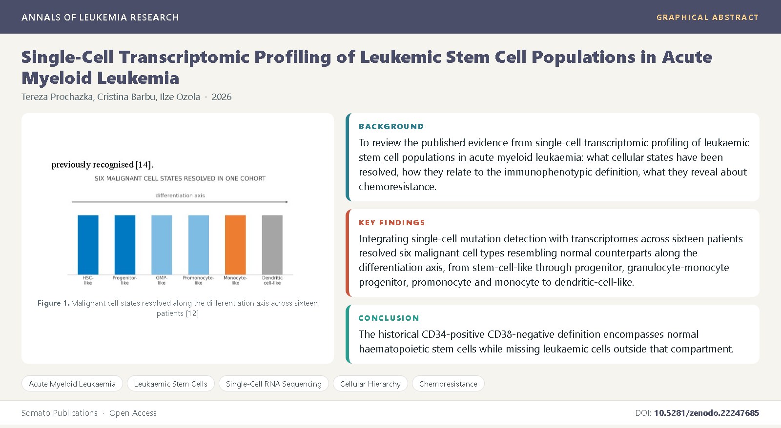
Leukaemic stem cells have been defined by an immunophenotype for nearly three decades. Single-cell sequencing shows that definition failing in both directions at once: it contains normal stem cells that should not be there, and misses leukaemic ones that lie outside the gate. Objectives: To review the published evidence from single-cell transcriptomic profiling of leukaemic stem cell populations in acute myeloid leukaemia: what cellular states have been resolved, how they relate to the immunophenotypic definition, what they reveal about chemoresistance, and whether the findings support a universal marker or a patient-specific one.
Material and Methods: Narrative review of single-cell sequencing studies, functional xenograft work, stemness score derivations and cellular hierarchy analyses indexed in PubMed and the major haematology and genomics journals. Studies were eligible if they characterised leukaemic stem or progenitor populations in acute myeloid leukaemia by single-cell methods, or established the phenotypic and functional definitions those methods have tested. No new patient data were generated and no patients were recruited; every finding quoted is one published by the original investigators. Results: Integrating single-cell mutation detection with transcriptomes across sixteen patients resolved six malignant cell types resembling normal counterparts along the differentiation axis, from stem-cell-like through progenitor, granulocyte-monocyte progenitor, promonocyte and monocyte to dendritic-cell-like, with
strikingly heterogeneous abundance between tumours: some comprised one or two types, others a full range. Leukaemic stem cells constitute under 10 per cent of malignant CD34-positive blasts. A quiescent immature stem-cell-like population was identified carrying raised AP-1 transcription factor expression and reduced class II major histocompatibility expression. A previously described surface marker was upregulated in only one of four patients examined. In thirteen paired pre- and post-chemotherapy paediatric samples, approximately half of leukaemic stem and progenitor populations actively expressed stem cell and oxidative phosphorylation signatures and showed enhanced metabolic programmes under treatment. Conclusion: The historical CD34-positive CD38-negative definition encompasses normal haematopoietic stem cells while missing leukaemic cells outside that compartment. Marker expression appears patient-specific rather than universal, which constrains what any single-marker targeting strategy can achieve.
Abbreviations: AML – Acute Myeloid Leukaemia, LSC – Leukaemic Stem Cell, HSC – Haematopoietic Stem Cell, LSPC – Leukaemic Stem and Progenitor Cell, scRNA-seq – Single-Cell RNA Sequencing, GMP – Granulocyte-Monocyte Progenitor, cDC – Conventional Dendritic Cell, OXPHOS – Oxidative Phosphorylation, MHC – Major Histocompatibility Complex, AP-1 – Activator Protein 1, MRD – Measurable Residual Disease, NOD/SCID – Non-Obese Diabetic Severe Combined Immunodeficiency.
References
Wu, J., Xiao, Y., Sun, J., Sun, H., Chen, H., Zhu, Y., et al. (2024) Unveiling novel insights in acute myeloid leukemia through single-cell RNA sequencing. Front Oncol, 14: 1365330.
Kantarjian, H., Kadia, T., DiNardo, C., Daver, N., Borthakur, G., Jabbour, E., et al. (2021) Acute myeloid leukemia: current progress and future directions. Blood Cancer J, 11: 41.
Döhner, H., Wei, AH., Appelbaum, FR., Craddock, C., DiNardo, CD., Dombret, H., et al. (2022) Diagnosis and management of AML in adults: 2022 recommendations from an international expert panel on behalf of the ELN. Blood, 140(12): 1345-1377.
Kayser, S., Levis, MJ. (2021) Updates on targeted therapies for acute myeloid leukaemia. Br J Haematol, 196(2): 316-328.
Bonnet, D., Dick, JE. (1997) Human acute myeloid leukemia is organized as a hierarchy that originates from a primitive hematopoietic cell. Nat Med, 3(7): 730-737.
Lapidot, T., Sirard, C., Vormoor, J., Murdoch, B., Hoang, T., Caceres-Cortes, J., et al. (1994) A cell initiating human acute myeloid leukaemia after transplantation into SCID mice. Nature, 367(6464): 645-648.
Roussel, X., Garnache-Ottou, F., Renosi, F. (2021) Rare classifications in acute leukemias and single-cell characterization. Cancers (Basel), 13(3): 505.
Hope, KJ., Jin, L., Dick, JE. (2004) Acute myeloid leukemia originates from a hierarchy of leukemic stem cell classes that differ in self-renewal capacity. Nat Immunol, 5(7): 738-743.
Pollyea, DA., Jordan, CT. (2017) Therapeutic targeting of acute myeloid leukemia stem cells. Blood, 129(12): 1627-1635.
Konopleva, MY., Jordan, CT. (2011) Leukemia stem cells and microenvironment: biology and therapeutic targeting. J Clin Oncol, 29(5): 591-599.
Wu, J., Xiao, Y., Sun, J., Sun, H., Chen, H., Zhu, Y., et al. (2020) A single-cell survey of cellular hierarchy in acute myeloid leukemia. J Hematol Oncol, 13(1): 128.
van Galen, P., Hovestadt, V., Wadsworth, MH II., Hughes, TK., Griffin, GK., Battaglia, S., et al. (2019) Single-cell RNA-Seq reveals AML hierarchies relevant to disease progression and immunity. Cell, 176(6): 1265-1281.e24.
Zeng, AGX., Bansal, S., Jin, L., Mitchell, A., Chen, WC., Abbas, HA., et al. (2022) A cellular hierarchy framework for understanding heterogeneity and predicting drug response in acute myeloid leukemia. Nat Med, 28(6): 1212-1223.
Yang, Y., Chen, X., Pan, J., Ning, H., Zhang, Y., Bo, Y., et al. (2026) Decoding the archipelago: single-cell biomarkers rechart the molecular geography of acute myeloid leukemia. Cell Commun Signal, 24: 88.
Jordan, CT., Upchurch, D., Szilvassy, SJ., Guzman, ML., Howard, DS., Pettigrew, AL., et al. (2000) The interleukin-3 receptor alpha chain is a unique marker for human acute myelogenous leukemia stem cells. Leukemia, 14(10): 1777-1784.
Bhatt, S., Pioso, MS., Olesinski, EA., Yilma, B., Ryan, JA., Mashaka, T., et al. (2024) Identification of leukemia stem cell subsets with distinct transcriptional, epigenetic and functional properties. Leukemia, 38(10): 2196-2209.
Ng, SWK., Mitchell, A., Kennedy, JA., Chen, WC., McLeod, J., Ibrahimova, N., et al. (2016) A 17-gene stemness score for rapid determination of risk in acute leukaemia. Nature, 540(7633): 433-437.
Velten, L., Story, BA., Hernandez-Malmierca, P., Raffel, S., Leonce, DR., Milbank, J., et al. (2021) Identification of leukemic and pre-leukemic stem cells by clonal tracking from single-cell transcriptomics. Nat Commun, 12(1): 1366.
Wu, J., Xiao, Y., Sun, J., Sun, H., Chen, H., Zhu, Y., et al. (2023) Single-cell transcriptomics reveals multiple chemoresistant properties in leukemic stem and progenitor cells in pediatric AML. Genome Biol, 24(1): 199.
Zeijlemaker, W., Grob, T., Meijer, R., Hanekamp, D., Kelder, A., Carbaat-Ham, JC., et al. (2019) CD34+CD38- leukemic stem cell frequency to predict outcome in acute myeloid leukemia. Leukemia, 33(5): 1102-1112.
Schuurhuis, GJ., Heuser, M., Freeman, S., Béné, MC., Buccisano, F., Cloos, J., et al. (2018) Minimal/measurable residual disease in AML: a consensus document from the European LeukemiaNet MRD Working Party. Blood, 131(12): 1275-1291.
Heuser, M., Freeman, SD., Ossenkoppele, GJ., Buccisano, F., Hourigan, CS., Ngai, LL., et al. (2021) 2021 update on MRD in acute myeloid leukemia: a consensus document from the European LeukemiaNet MRD Working Party. Blood, 138(26): 2753-2767.
Petti, AA., Williams, SR., Miller, CA., Fiddes, IT., Srivatsan, SN., Chen, DY., et al. (2019) A general approach for detecting expressed mutations in AML cells using single cell RNA-sequencing. Nat Commun, 10(1): 3660.
Ng, SW., Murphy, T., King, I., Zhang, T., Mah, M., Lu, Z., et al. (2022) A clinical laboratory-developed LSC17 stemness score assay for rapid risk assessment of acute myeloid leukemia patients. Blood Adv, 6(4): 1064-1073.
Boutzen, H., Madani Tonekaboni, SA., Chan-Seng-Yue, M., Murison, A., Takayama, N., Mbong, N., et al. (2022) A primary hierarchically organized patient-derived model enables in depth interrogation of stemness driven by the coding and non-coding genome. Leukemia, 36(5): 1421-1425.
Papaemmanuil, E., Gerstung, M., Bullinger, L., Gaidzik, VI., Paschka, P., Roberts, ND., et al. (2016) Genomic classification and prognosis in acute myeloid leukemia. N Engl J Med, 374(23): 2209-2221.
Shlush, LI., Mitchell, A., Heisler, L., Abelson, S., Ng, SWK., Trotman-Grant, A., et al. (2017) Tracing the origins of relapse in acute myeloid leukaemia to stem cells. Nature, 547(7661): 104-108.
Jan, M., Snyder, TM., Corces-Zimmerman, MR., Vyas, P., Weissman, IL., Quake, SR., Majeti, R. (2012) Clonal evolution of preleukemic hematopoietic stem cells precedes human acute myeloid leukemia. Sci Transl Med, 4(149): 149ra118.
Corces-Zimmerman, MR., Hong, WJ., Weissman, IL., Medeiros, BC., Majeti, R. (2014) Preleukemic mutations in human acute myeloid leukemia affect epigenetic regulators and persist in remission. Proc Natl Acad Sci USA, 111(7): 2548-2553.
Farge, T., Saland, E., de Toni, F., Aroua, N., Hosseini, M., Perry, R., et al. (2017) Chemotherapy-resistant human acute myeloid leukemia cells are not enriched for leukemic stem cells but require oxidative metabolism. Cancer Discov, 7(7): 716-735.
Lagadinou, ED., Sach, A., Callahan, K., Rossi, RM., Neering, SJ., Minhajuddin, M., et al. (2013) BCL-2 inhibition targets oxidative phosphorylation and selectively eradicates quiescent human leukemia stem cells. Cell Stem Cell, 12(3): 329-341.
Ishikawa, F., Yoshida, S., Saito, Y., Hijikata, A., Kitamura, H., Tanaka, S., et al. (2007) Chemotherapy-resistant human AML stem cells home to and engraft within the bone-marrow endosteal region. Nat Biotechnol, 25(11): 1315-1321.