Differential Radiology Diagnostics of Rare Cystic Pancreatic Lesions
Keywords:
Cystic pancreatic neoplasm, US, MSCT, MRI, Radiology, IPMN, SPPT, DiagnosticsAbstract
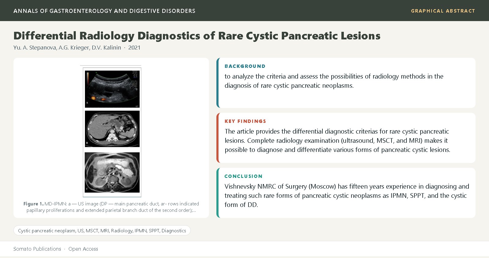
The purpose: to analyze the criteria and assess the possibilities of radiology methods in the diagnosis of rare cystic pancreatic neoplasms. Materials and methods: From 2004 to 2019 at A.V. Vishnevsky NMRC of Surgery 222 patients with suspected cystic pancreatic lesions were examined and treated, 15-80 years old, men - 59.9%. Radiology examination: ultrasound, multislice computed tomography (MSCT) and magnetic resonance imaging (MRI). 182 patients (82.0%) were operated. Morphology: intraductal papillary mucinous neoplasms (IPMN) - 96 (43.2%), solid pseudopapillary tumors (SPPT) - 50 (22.6%), cystic form of duodenal dystrophy (DD) - 72 (32.4%), cystic teratoma - 1 (0.45%), cystic lymphangioma – 1 (0.45%), echinococcosis – 2 cases (0.9%). Results: The article provides the differential diagnostic criterias for rare cystic pancreatic lesions. Complete radiology examination (ultrasound, MSCT, and MRI) makes it possible to diagnose and differentiate various forms of pancreatic cystic lesions. At suspected presence of a rare pancreatic lesion, the radiology preference should be given to MRI. Criteria for radiology diagnostics of the IPMN and DD cystic form have been well defined. The formulation of the SPPT diagnostic criteria requires the accumulation of a greater number of observations in one place and an analysis of the corresponding information. Anamnesis of the disease is an important supplement to the SPPT radiology diagnostics. Conclusions: A.V. Vishnevsky NMRC of Surgery (Moscow) has fifteen years experience in diagnosing and treating such rare forms of pancreatic cystic neoplasms as IPMN, SPPT, and the cystic form of DD. It should be pointed
References
The European Study Group on Cystic Tumours of the Pancreas (2018) European evidence-based guidelines on pancreatic cystic neoplasms. Gut, 67: 789–804.
Le Borgne, J., de Calan, L., Partensky, C. (1999) Cystadenomas and cystadenocarcinomas of the pancreas: a multiinstitutional retrospective study of 398 cases. French Surgical Association. Ann Surg, 230(2): 152-161.
Lim, JH., Lee, G., Oh, YL. (2001) Radiologic Spectrum of Intraductal Papillary Mucinous Tumor of the Pancreas. Radiographics, 21: 323–337.
Ohhashi, K., Murakami, F., Maruyama, M., Takekoshi, T., Ohta, H., Ohhashi, I., et al. (1982) Four cases of mucous secreting pancreatic cancer. Prog Digest Endosc, 20: 348– 351.
Stepanova, YA., Karmazanovsky, GG., Egorov, VI., Kochatkov, AV., Dubova, EA., Kosova, IA., et al. (2009) Radiology methods of the diagnosis of intraductal papillary mucinous neoplasms. Annals of Surgical Hepatology, 14(3): 69-79.
The European Study Group on Cystic Tumours of the Pancreas (2018) European evidence-based guidelines on pancreatic cystic neoplasms. Gut, 67: 789–804.
Frantz, VK. (1959) Tumors of the pancreas: Atlas of tumor pathology. US Armed Forces Institute of pathology. Washington DC, 32-33.
Klöppel, G., Solcia, E., Longnecker, DS., Capella, C., Les - lie, HS. in collaboration with pathologists in 7 countries (1996) Histological Typing of Tumours of the Exocrine Pancreas (WHO. World Health Organization. International Histological Classification of Tumours). 2nd ed. Berlin: Springer-Verlag, 72p.
World Health Organization classification of tumours (2000) Pathology and genetics of tumours of the digestive system. Hamilton S.R., Altonen L.A. Eds. Lyon: IARC Press, 314p.
Papavramidis, T., Papavramidis, S. (2005) Solid pseudopapillary tumors of the pancreas: review of 718 patients reported in English literature. J Am Coll Surg, 200(6): 965-972.
Vassos, N., Agaimy, A., Klein, P., Hohenberger, W., Croner, RS. (2013) Solid-pseudopapillary neoplasm (SPN) of the pancreas: case series and literature review on an enigmatic entity. Int J Clin Exp Pathol, 6(6): 1051-1059.
Shiralkar, M., Adler, DG. (2020) Solid Pseudopapillary Tumors of the Pancreas: A Rare but Important Clinical Entity. Practical gastro, XLIV(2): 22-29.
Laje, P., Bhatti, TR., Adzick, NS. (2013) Solid pseudopapillary neoplasm of the pancreas in children: a 15-year experience and the identification of a unique immunohistochemical marker. J Pediatr Surg, 48(10): 2054-2060.
Yu, CC., Tseng, JH., Yeh, CN., Hwang, TL., Jan, YY. (2007) Clinicopathological study of solid and pseudopapillary tumor of pancreas: emphasis on magnetic resonance imaging findings. World J Gastroenterol, 13(12): 1811-1815.
Zinner, MJ., Shurbaji, MS., Cameron, JL. (1990) Solid and papillary epithelial neoplasms of the pancreas. Surgery, 108(3): 475-480.
Eder, F., Schulz, HU., Röcken, C., Lippert, H. (2005) Solid-pseudopapillary tumor of the pancreatic tail. World J Gastroenterol, 11(26): 4117-4119.
Antoniou, EA., Damaskos, C., Garmpis, N., Salakos, C., Margonis, GA., Kontzoglou K., Lahanis, S., et al. (2017) Solid Pseudopapillary Tumor of the Pancreas: A Single-center Experience and Review of the Literature. In Vivo, 31(4): 501-510.
Salvia, R., Bassi, C., Festa, L., Falconi, M., Crippa, S., Butturini, G., et al. (2007) Clinical and biological behavior of pancreatic solid pseudopapillary tumors: report on 31 consecutive patients. J Surg Oncol, 95(4): 304-310.
Nishihara, K., Nagoshi, M., Tsuneyoshi, M., Yamaguchi, K., Hayashi, I. (1993) Papillary cystic tumors of the pancreas. Assessment of their malignant potential. Cancer, 71(1): 82-92.
Song, H., Dong, M., Zhou, J., Sheng, W., Zhong, B., Gao, W. (2017) Solid Pseudopapillary Neoplasm of the Pancreas: Clinicopathologic Feature, Risk Factors of Malignancy, and Survival Analysis of 53 Cases from a Single Center. Biomed Res Int, 5465261.
Asano, T., Seya, T., Tanaka, N., Ooaki, Y., Fujino, O. (2006) A 13-year-old girl with a preoperatively diagnosed solid cystic tumor of the pancreas. J Nippon Med Sch, 73(4): 231-234.
Dolan, RV., ReMine, WH., Dockerty, MB. (1974) The fate of heterotopic pancreatic tissue. A study of 212 cases. Arch Surg, 109(6): 762-765.
Skandalakis, JE., Grey, SW. (1994) Embryology for surgeons: the embryological basis for treatment of congenital anomalies, 2nd ed., Baltimore (MD), USA: Williams Wilkins, 366-387.
Skandalakis, JE., Skandalakis, LJ., Colborn, GL. (1998) Congenital anomalies and variations of the pancreas and pancreatic and extrahepatic bile ducts. In: The Pancreas, H.G. Beger et al. ed., Oxford: Blackwell Science, 28-30.
Gabata, T., Kadoya, M., Terayama, N., Sanada, J., Kobayashi, S., Matsui, O. (2003) Groove pancreatic carcinomas: radiological and pathological findings. European Radiology, 13(7): 1679-1684.
Potet, F., Duclert, N. (1970) Dystrophie kystique sur pancreas aberrant de la paroi duodenale. Arch Fr Mal App Dig, 59: 223.
Freeny, PC. (1998) Radiology. In: The Pancreas. H.G. Beger et al. ed., Oxford: Blackwell Science, 1024.
Frulloni, L., Gabbrielli, A., Pezzilli, R., Zerbi, A., Cavestro, GM., Marotta, F., et al. (2009) Chronic pancreatitis: report from a multicenter Italian survey (PanCroInfAISP) on 893 patients. Dig Liver Dis, 41(4): 311-317.
Koch, K. (1913) Beiträge zur Pathologie der Bauchspeicheldrüse. Virchows Archiv, 214(2): 180–206.
Lyngdoh, TS., Konsam, R., Th, B., Marak, B. (2008) Giant cystic lymphangioma of the pancreas. ANZ J Surg, 78(8): 673-674.
Fonseca, R., Pitman, MB. (2013) Lymphangioma of the pancreas: a multimodal approach to pre-operative diagnosis. Cytopathology, 24(3): 172-176.
Stepanova, YA., Karmazanovsky, GG., Kubyshkin, VA., Schegolev, AI. (2013) Rare Cystic Masses of the Pancreas: Differential Radiological Diagnosis. Ukrainian Journal of Surgery, 3(22): 99-115.
Kerr, AA. (1922) Cysts and pseudocysts of the pancreas. Surg Gynecol Obstet, 27: 40.
Primrose, A. (1922) Pancreatic cyst and pseudocyst. Surg Gynecol Obstet, 34: 431-436.
Zhi, L., Nengwen, K., Xubao, L., Shu, G. (2018) Mature cystic teratoma of the pancreas with 30 years of clinical course. A case report. Medicine, 97(15): e0405.
Chakaravarty, KD., Venkata, CD., Manicketh, I., Singh, R., Mathew, P., Devashetty, S., et al. (2016) Mature Cystic Teratoma of the Pancreas. ACG Case Rep J, 3(2): 80-81.
Makni, A., Jouini, M., Kacem, M., Safta, ZB. (2012) Acute pancreatitis due to pancreatic hydatid cyst: a case report and review of the literature. World J Emerg Surg, 7(1): 7.
Soin, P., Sharma, P., Kochar, PS. (2019) Pancreatic echinococcosis. Proc (Bayl Univ Med Cent)., 32(1): 85-87.
Ahmed, Z., Chhabra, S., Massey, A., Vij, V., Yadav, R., Bugalia, R., et al. (2016) Primary hydatid cyst of pancreas: Case report and review of literature. Int J Surg Case Rep, 27: 74-77.
Dahniya, MH., Hanna, RM., Ashebu, S., Muhtaseb SA., el-Beltagi, A., Badr, S., et al. (2001) The imaging appearances of hydatid disease at some unusual sites. Br J Radi (879): 283-289.
Kattan, YB. (1975) Hydatid cysts in pancreas. Br Med J, 4(5999): 729-730.
Ozmen, MM., Moran, M., Karakahya, M., Coskun, F. (2005) Recurrent acute pancreatitis due to a hydatid cyst of the pancreatic head: a case report and review of the literature. JOP, 6(4): 354-358.