Cerebral Calcifications in Systemic Lupus Erythematosus
Abstract
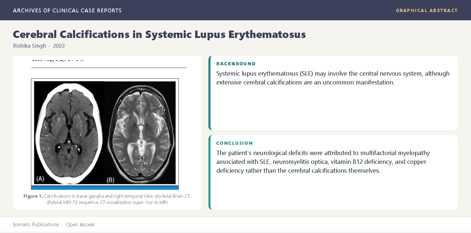
Background: Systemic lupus erythematosus (SLE) may involve the central nervous system, although extensive cerebral calcifications are an uncommon manifestation. Intracranial calcifications can result from various etiologies and are more accurately detected by computed tomography (CT) than magnetic resonance imaging (MRI). Case Presentation: A 43-year-old African American woman with a 13-year history of SLE presented with progressive right leg weakness, urinary incontinence, and recurrent falls. Neuroimaging revealed extensive bilateral cerebral calcifications involving the basal ganglia, cerebellum, brainstem, and temporal lobe. MRI additionally demonstrated chronic small-vessel ischemic changes and cervicothoracic spinal cord abnormalities. Laboratory investigations showed elevated inflammatory markers, positive aquaporin-4 antibodies, and cerebrospinal fluid findings consistent with central nervous system inflammation. Differential diagnoses, including metabolic disorders and Fahr’s disease, were considered and excluded based on clinical and laboratory findings. Discussion: The patient's neurological deficits were attributed to multifactorial myelopathy associated with SLE, neuromyelitis optica, vitamin B12 deficiency, and copper deficiency rather than the cerebral calcifications themselves. The intracranial calcifications were considered an incidental manifestation of cerebral lupus. This case highlights the importance of correlating imaging findings with clinical presentation and recognizing that extensive cerebral calcifications in SLE do not necessarily account for neurological symptoms. Conclusion: Cerebral calcifications are a rare but recognized feature of central nervous system involvement in SLE and are best identified using CT imaging. Comprehensive clinical assessment is essential to distinguish incidental radiological findings from the true cause of neurological deficits, enabling appropriate diagnosis and management.
References
Yamada, M., Asano, T., Kouichirou, O., Yuichi, H., Masayuki, K., Hiroaki, H., et al. (2013) High frequency of calcification in basal ganglia on brain computed tomography images in Japanese older adults. Geriatr Gerontol Int, 13(3): 706- 710.
Saade, C., Najem, E., Asmar, K., Salman, R., El-Achkar, B., Naffaa, L. (2019) Intracranial calcifications on CT: an updated review. J Radiol Case Rep, 13(8):1-18.
NORD 2022; https://rarediseases.org/rare-diseases/primary-familial-brain-calcification.
Martinovic-Kaliterna, D., Radic, M., Radic, J., Kovacic, V., Fabijanic, D. (2013) Massive cerebral calcifications (Fahr’s disease) in a patient with systemic lupus erythematosus and no major neuropsychological abnormality. Isr Med Assoc J, 15(10): 654-655.
Joseph, FG., Lammie, GA., Scolding, NJ. (2007) CNS lupus: A study of 41 patients. Neurology, 69(7): 644-654.
Raymond, AA., Zariah, AA., Samad, SA., Chin, CN., Kong, NCT. (1996) Brain calcification in patients with cerebral lupus. Lupus, 5(2): 123-128.
Manyam, BV. (2005) What is and what is not “Fahr’s disease.” Parkinsonism and Related Disorders, 11(2): 73-80.