skin barrier function testing in the Management of facial hyperpigmentation among adult dermatology patients
DOI:
https://doi.org/10.5281/zenodo.22814457Keywords:
skin barrier function, facial hyperpigmentation, melasma, transepidermal water loss, stratum corneumAbstract
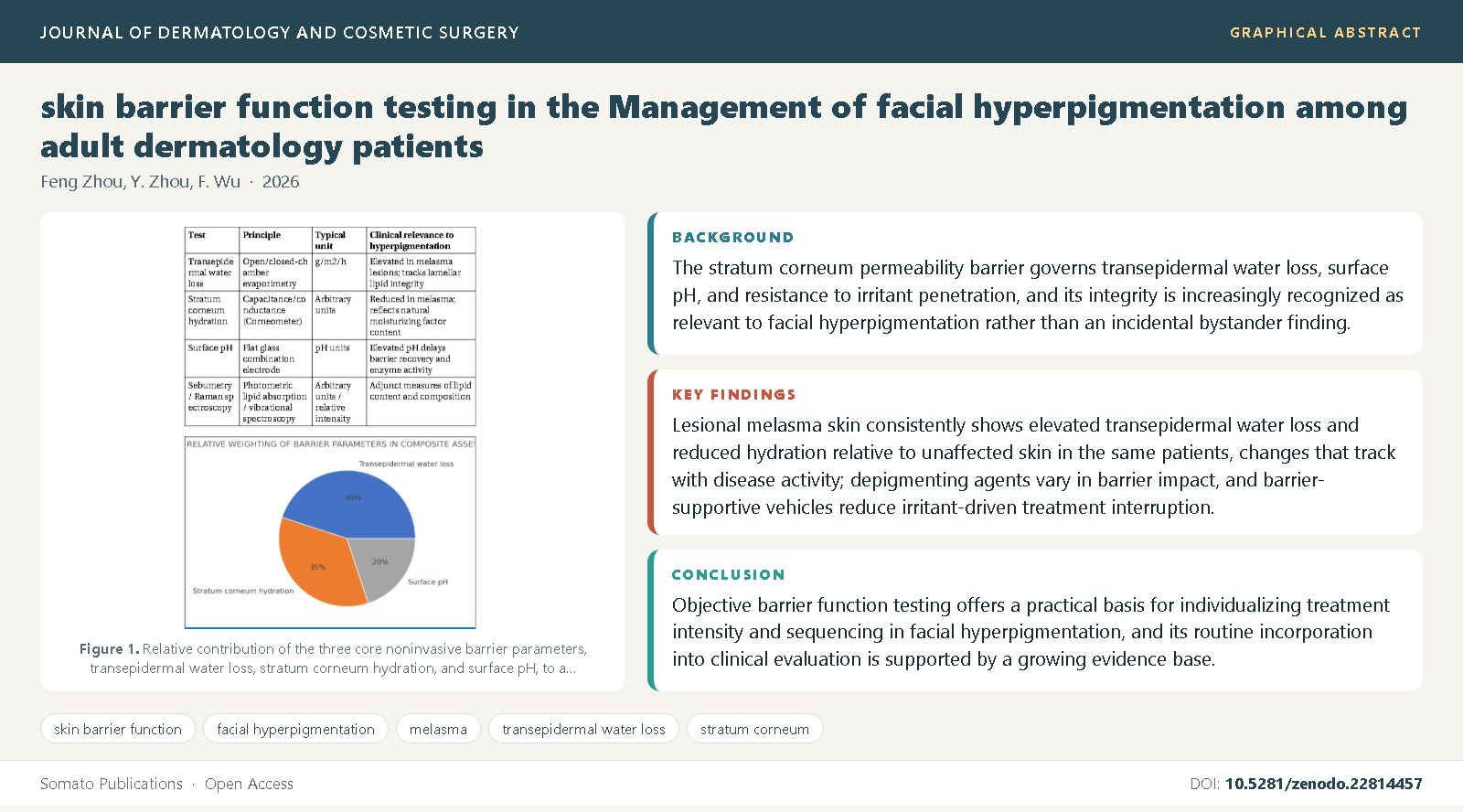
Background: The stratum corneum permeability barrier governs transepidermal water loss, surface pH, and resistance to irritant penetration, and its integrity is increasingly recognized as relevant to facial hyperpigmentation rather than an incidental bystander finding.
Objective: This review synthesizes evidence on noninvasive skin barrier function testing in adult patients with facial hyperpigmentation, principally melasma, and how barrier assessment can inform treatment selection.
Methods: We synthesized comparative biophysical studies, methodological guidelines, and treatment trials addressing transepidermal water loss, hydration, surface pH, and their relationship to pigmentary disease activity and tolerability.
Findings: Lesional melasma skin consistently shows elevated transepidermal water loss and reduced hydration relative to unaffected skin in the same patients, changes that track with disease activity; depigmenting agents vary in barrier impact, and barrier-supportive vehicles reduce irritant-driven treatment interruption.
Conclusions: Objective barrier function testing offers a practical basis for individualizing treatment intensity and sequencing in facial hyperpigmentation, and its routine incorporation into clinical evaluation is supported by a growing evidence base.
References
Elias, P.M. (2005) Stratum corneum defensive functions: an integrated view. Journal of Investigative Dermatology, 125(2): 183-200. DOI: 10.1111/j.0022-202X.2005.23668.x
Madison, K.C. (2003) Barrier function of the skin: "la raison d'etre" of the epidermis. Journal of Investigative Dermatology, 121(2): 231-241. DOI: 10.1046/j.1523-1747.2003.12359.x
Kligman, A.M. (1978) Perspectives and problems in cutaneous gerontology. Journal of Investigative Dermatology, 73(1): 39-46. DOI: 10.1111/1523-1747.ep12532758
Pinnagoda, J., Tupker, R.A., Agner, T., Serup, J. (1990) Guidelines for transepidermal water loss (TEWL) measurement. Contact Dermatitis, 22(3): 164-178. DOI: 10.1111/j.1600-0536.1990.tb01553.x
Berardesca, E., Loden, M., Serup, J., Masson, P., Rodrigues, L.M. (2018) The revised EEMCO guidance for the in vivo measurement of water in the skin. Skin Research and Technology, 24(3): 351-358. DOI: 10.1111/srt.12599
Darlenski, R., Sassning, S., Tsankov, N., Fluhr, J.W. (2013) Non-invasive in vivo methods for investigation of the skin barrier physical properties. European Journal of Pharmaceutics and Biopharmaceutics, 72(2): 295-303. DOI: 10.1016/j.ejpb.2008.11.013
Lee, A.Y. (2015) Recent progress in melasma pathogenesis. Pigment Cell and Melanoma Research, 28(6): 648-660. DOI: 10.1111/pcmr.12404
Kwon, S.H., Hwang, Y.J., Lee, S.K., Park, K.C. (2016) Heterogeneous pathology of melasma and its clinical implications. International Journal of Molecular Sciences, 17(6): 824. DOI: 10.3390/ijms17060824
Lee, D.J., Park, K.C., Ortonne, J.P., Kang, H.Y. (2012) Pendulous melanocytosis: a reconsideration of the histopathology of solar lentigo and melasma. Pigment Cell and Melanoma Research, 25(1): 1-6. DOI: 10.1111/j.1755-148X.2011.00913.x
Regazzetti, C., De Donatis, G.M., Ghorbel, H.H., Cardot-Leccia, N., Ambrosetti, D., Bahadoran, P., et al. (2015) Endothelial cells promote pigmentation through endothelin receptor B activation. Journal of Investigative Dermatology, 135(12): 3096-3104. DOI: 10.1038/jid.2015.332
Fluhr, J.W., Feingold, K.R., Elias, P.M. (2006) Transepidermal water loss reflects permeability barrier status: validation in human and rodent in vivo and ex vivo models. Experimental Dermatology, 15(7): 483-492. DOI: 10.1111/j.1600-0625.2006.00437.x
Man, M.Q., Xin, S.J., Song, S.P., Cho, S.Y., Zhang, X.J., Tu, C.X., et al. (2009) Variation of skin surface pH, sebum content and stratum corneum hydration with age and gender in a large Chinese population. Skin Pharmacology and Physiology, 22(4): 190-199. DOI: 10.1159/000231524
Ghadially, R., Brown, B.E., Sequeira-Martin, S.M., Feingold, K.R., Elias, P.M. (1995) The aged epidermal permeability barrier: structural, functional, and lipid biochemical abnormalities in humans and a senescent murine model. Journal of Clinical Investigation, 95(5): 2281-2290. DOI: 10.1172/JCI117919
Nikolovski, J., Stamatas, G.N., Kollias, N., Wiegand, B.C. (2008) Barrier function and water-holding and transport properties of infant stratum corneum are different from adult and continue to develop through the first year of life. Journal of Investigative Dermatology, 128(7): 1728-1736. DOI: 10.1038/sj.jid.5701239
Wan, D.C., Wong, V.W., Longaker, M.T., Yang, G.P., Wei, F.C. (2014) Moisturizing different racial skin types. Journal of Clinical and Aesthetic Dermatology, 7(6): 25-32.
Alexander, H., Brown, S., Danby, S., Flohr, C. (2018) Research techniques made simple: transepidermal water loss measurement as a research tool. Journal of Investigative Dermatology, 138(11): 2295-2300. DOI: 10.1016/j.jid.2018.09.001
Eberlein-Konig, B., Schafer, T., Huss-Marp, J., Darsow, U., Mohrenschlager, M., Herbert, O., et al. (2000) Skin surface pH, stratum corneum hydration, trans-epidermal water loss and skin roughness related to atopic eczema and skin dryness in a population of primary school children. Acta Dermato-Venereologica, 80(3): 188-191. DOI: 10.1080/000155500750042943
Yin, N.C., Wang, S.Q. (2017) Skin barrier function: how does it work and can it be quantified in humans? Journal of Cosmetic Dermatology, 16(3): 351-356. DOI: 10.1111/jocd.12336
Lee, D.J., Lee, J., Ha, J., Park, K.C., Ortonne, J.P., Kang, H.Y. (2009) Defective barrier function in melasma skin. Journal of the European Academy of Dermatology and Venereology, 23(12): 1400-1404. DOI: 10.1111/j.1468-3083.2009.03323.x
Jeon, S.Y., Pyo, H.K., Kim, W.S. (2016) Comparison of skin barrier function in melasma and normal skin. Journal of Cosmetic Dermatology, 15(4): 465-470. DOI: 10.1111/jocd.12235
Handa, S., Kaur, R., De, D., Mahajan, R., Sethi, S., Sharma, N. (2019) Sequential biophysical characterization of melasma over time. International Journal of Dermatology, 58(2): 214-219. DOI: 10.1111/ijd.14197
Huang, C.T., Chen, C.L., Liao, C.C., Chen, L.Y., Tsai, J.C. (2020) Skin barrier function analysis and correlation with dermoscopy in patients with melasma. International Journal of Dermatology, 59(4): 456-462. DOI: 10.1111/ijd.14741
Kim, N.H., Lee, C.H., Lee, A.Y. (2007) Cadherins, an important mediator of keratinocyte-melanocyte interactions in the epidermis. Journal of Investigative Dermatology, 127(3): 693-702. DOI: 10.1038/sj.jid.5700580
Seiberg, M. (2001) Keratinocyte-melanocyte interactions during melanosome transfer. Pigment Cell Research, 14(4): 236-242. DOI: 10.1034/j.1600-0749.2001.140402.x
Yamaguchi, Y., Hearing, V.J. (2009) Physiological factors that regulate skin pigmentation. BioFactors, 35(2): 193-199. DOI: 10.1002/biof.29
Costin, G.E., Hearing, V.J. (2007) Human skin pigmentation: melanocytes modulate skin color in response to stress. FASEB Journal, 21(4): 976-994. DOI: 10.1096/fj.06-6649rev
Ortonne, J.P., Arellano, I., Berneburg, M., Cestari, T., Chan, H., Grimes, P., et al. (2009) A global survey of the role of ultraviolet radiation and hormonal influences in the development of melasma. Journal of the European Academy of Dermatology and Venereology, 23(11): 1254-1262. DOI: 10.1111/j.1468-3083.2009.03295.x
Gilchrest, B.A. (2013) Photoaging. Journal of Investigative Dermatology, 133(E1): E2-E6. DOI: 10.1038/skinbio.2013.176
Passeron, T., Picardo, M. (2018) Melasma, a photoaging disorder. Pigment Cell and Melanoma Research, 31(4): 461-465. DOI: 10.1111/pcmr.12684
Gonzalez, S., Fernandez-Lorente, M., Gilaberte-Calzada, Y. (2008) The latest on skin photoprotection. Clinics in Dermatology, 26(6): 614-626. DOI: 10.1016/j.clindermatol.2007.09.010
Seite, S., Bieber, T. (2015) Barrier function and microbiotic dysbiosis in atopic dermatitis. Clinical, Cosmetic and Investigational Dermatology, 8: 479-483. DOI: 10.2147/CCID.S91521
Callender, V.D., St Surin-Lord, S., Davis, E.C., Maclin, M. (2011) Postinflammatory hyperpigmentation: etiologic and therapeutic considerations. American Journal of Clinical Dermatology, 12(2): 87-99. DOI: 10.2165/11536930-000000000-00000
Sarkar, R., Arora, P., Garg, K.V. (2013) Cosmeceuticals for hyperpigmentation: what is available? Journal of Cutaneous and Aesthetic Surgery, 6(1): 4-11. DOI: 10.4103/0974-2077.110089
Rendon, M., Berneburg, M., Arellano, I., Picardo, M. (2006) Treatment of melasma. Journal of the American Academy of Dermatology, 54(5 Suppl 2): S272-S281. DOI: 10.1016/j.jaad.2005.12.043
Sheth, V.M., Pandya, A.G. (2011) Melasma: a comprehensive update: part I. Journal of the American Academy of Dermatology, 65(4): 689-697. DOI: 10.1016/j.jaad.2010.12.046
Choi, Y.J., Kim, J.H., Chung, H.C., Choi, H.J., Chung, W.G., Cho, K.H., et al. (2018) A comparative study of the efficacy of tranexamic acid and vitamin C in melasma with skin barrier assessment. Annals of Dermatology, 30(6): 733-735. DOI: 10.5021/ad.2018.30.6.733
Lima, P.B., Dias, J.A.F., Cassiano, D., Esposito, A.C.C., Miot, L.D.B., Bagatin, E., et al. (2020) A comparative study of topical 5% cysteamine versus 4% hydroquinone in the treatment of facial melasma in women. International Journal of Dermatology, 59(10): 1245-1250. DOI: 10.1111/ijd.15082
Grimes, P.E., Ijaz, S., Nashawati, R., Kwak, D. (2019) New oral and topical approaches for the treatment of melasma. International Journal of Women's Dermatology, 5(1): 30-36. DOI: 10.1016/j.ijwd.2018.09.004
Loden, M. (2003) Role of topical emollients and moisturizers in the treatment of dry skin barrier disorders. American Journal of Clinical Dermatology, 4(11): 771-788. DOI: 10.2165/00128071-200304110-00005

Published
Issue
Section
License
Copyright (c) 2026 Feng Zhou, et al.

This work is licensed under a Creative Commons Attribution 4.0 International License.