Evaluation of dermoscopic evaluation as a Prognostic Tool in plaque psoriasis
DOI:
https://doi.org/10.5281/zenodo.22814352Keywords:
dermoscopy, plaque psoriasis, prognosis, vascular pattern, treatment responseAbstract
Background: Chronic plaque psoriasis is diagnosed and monitored largely by unaided inspection, an approach that performs poorly for thin plaques, darker phototypes, and distinguishing psoriasis from its papulosquamous mimics.
Objective: This review evaluates the evidence on dermoscopic examination of plaque psoriasis as a prognostic tool, covering its correlation with severity, its use in monitoring biologic therapy response, and its role in differential diagnosis.
Methods: We synthesized diagnostic-accuracy studies, cross-sectional severity-correlation cohorts, and serial imaging studies addressing dermoscopic vascular and scale patterns in chronic plaque psoriasis.
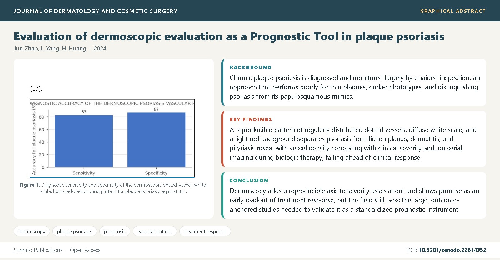
Findings: A reproducible pattern of regularly distributed dotted vessels, diffuse white scale, and a light red background separates psoriasis from lichen planus, dermatitis, and pityriasis rosea, with vessel density correlating with clinical severity and, on serial imaging during biologic therapy, falling ahead of clinical response.
Conclusions: Dermoscopy adds a reproducible axis to severity assessment and shows promise as an early readout of treatment response, but the field still lacks the large, outcome-anchored studies needed to validate it as a standardized prognostic instrument.
References
Lallas, A., Kyrgidis, A., Tzellos, T.G., Apalla, Z., Karakyriou, E., Karatolias, A., et al. (2013) Accuracy of dermoscopic criteria for the diagnosis of psoriasis, dermatitis, lichen planus and pityriasis rosea. British Journal of Dermatology, 168(3): 616-624. DOI: 10.1111/bjd.12196
Vazquez-Lopez, F., Manjon-Haces, J.A., Maojo-Fernandez, C., Guerra-Tapia, A., Perez-Oliva, N., Marghoob, A.A. (2004) Dermoscopic features of plaque psoriasis and lichen planus: new observations. Dermatology, 207(2): 151-156. DOI: 10.1159/000079668
Parisi, R., Symmons, D.P.M., Griffiths, C.E.M., Ashcroft, D.M. (2013) Global epidemiology of psoriasis: a systematic review of incidence and prevalence. Journal of Investigative Dermatology, 133(2): 377-385. DOI: 10.1038/jid.2012.339
Errichetti, E., Piccirillo, A., Stinco, G. (2016) Dermoscopy of psoriasis: an updated review. Giornale Italiano di Dermatologia e Venereologia, 151(4): 411-419.
Lacarrubba, F., Pellacani, G., Verzi, A.E., Pippione, M., Micali, G. (2015) Dermoscopy in the diagnosis and management of chronic plaque psoriasis: a review. Dermatology Practical & Conceptual, 5(3): 5-11. DOI: 10.5826/dpc.0503a02
Russo, T., Piccolo, V., Lallas, A., Argenziano, G. (2015) Recent advances in dermoscopy. F1000Research, 5: 1712. DOI: 10.12688/f1000research.8801.1
Griffiths, C.E.M., Barker, J.N.W.N. (2007) Pathogenesis and clinical features of psoriasis. Lancet, 370(9583): 263-271. DOI: 10.1016/S0140-6736(07)61128-3
Heidenreich, R., Rocken, M., Ghoreschi, K. (2009) Angiogenesis drives psoriasis pathogenesis. International Journal of Experimental Pathology, 90(3): 232-248. DOI: 10.1111/j.1365-2613.2009.00669.x
Vazquez-Lopez, F., Alvarez-Cuesta, C., Hidalgo-Garcia, Y., Perez-Oliva, N. (2003) The handheld dermoscope improves the recognition of the specific dotted and globular vessels of plaque psoriasis. Archives of Dermatology, 139(8): 1078. DOI:
1001/archderm.139.8.1078 10. Errichetti, E., Zalaudek, I., Kittler, H., Apalla, Z., Argenziano, G., Bakos, R., et al. (2017) Standardization of dermoscopic terminology and basic dermoscopic parameters to evaluate in general dermatology (non-neoplastic dermatoses): an expert consensus on behalf of the International Dermoscopy Society. British Journal of Dermatology, 182(2): 454-467. DOI: 10.1111/bjd.18125
Zalaudek, I., Argenziano, G. (2010) Dermoscopy subpatterns of inflammatory skin disorders. Archives of Dermatology, 146(10): 1182. DOI: 10.1001/archdermatol.2010.288
Kaczorowska, A., Rudnicka, L., Szczepanik-Kulak, P., Warszawik-Hendzel, O. (2020) Dermoscopy of nonspecific inflammatory dermatoses. Dermatology and Therapy, 10(4): 615-624. DOI: 10.1007/s13555-020-00378-y
Kelati, A., Aqil, N., Baybay, H., Gallouj, S., Mernissi, F.Z. (2017) Dermoscopy: a useful tool for diagnosing palmoplantar psoriasis in the darker phototype. International Journal of Dermatology, 56(3): 314-320. DOI: 10.1111/ijd.13564
Gutte, R., Khopkar, U. (2016) Dermoscopy of nail psoriasis. Indian Journal of Dermatology, Venereology and Leprology, 82(1): 22-24. DOI: 10.4103/0378-6323.171990
Errichetti, E., Piccirillo, A., Viola, L., Stinco, G. (2015) Dermatoscopy of extra-facial and facial seborrheic dermatitis compared with psoriasis. Journal of the American Academy of Dermatology, 73(1): e21-e22. DOI: 10.1016/j.jaad.2015.03.012
Lallas, A., Argenziano, G., Apalla, Z., Gourhant, J.Y., Zaballos, P., Di Lernia, V., et al. (2012) Dermoscopic patterns of common facial inflammatory skin diseases. Journal of the European Academy of Dermatology and Venereology, 26(8): 1017-1021. DOI: 10.1111/j.1468-3083.2011.04440.x
Pan, Y., Chamlin, S.L., Cordoro, K.M. (2020) Dermoscopic vascular patterns in pediatric psoriasis and their correlation with disease severity. Pediatric Dermatology, 37(6): 1055-1060. DOI: 10.1111/pde.14345
Gisondi, P., Geat, D., Nald, R.D., Girolomoni, G. (2020) Reliability of clinical scores and dermoscopy in assessing severity of chronic plaque psoriasis. Journal of the European Academy of Dermatology and Venereology, 34(8): 1786-1791. DOI: 10.1111/jdv.16223
Shobana, K., Gowda, N.C., Sudhir, N.U., Kishore, B.N., Basavaraj, V. (2019) Dermoscopic evaluation of psoriasis and its correlation with severity. Indian Dermatology Online Journal, 10(4): 421-425. DOI: 10.4103/idoj.IDOJ_338_18
Feldman, S.R., Fleischer, A.B., Reboussin, D.M., Rice, J.P., Andrews, K., Rajadhyaksha, V., et al. (1996) The self-administered psoriasis area and severity index is valid and reliable. Journal of Investigative Dermatology, 106(1): 183-186. DOI: 10.1111/1523-1747.ep12329912
Schmitt, J.M., Ford, D.E. (2005) Reliability and validity of the psoriasis area and severity index (PASI) in patients with moderate to severe psoriasis: a critical review. British Journal of Dermatology, 152(5): 861-867. DOI: 10.1111/j.1365-2133.2005.06503.x
Langley, R.G.B., Ellis, C.N. (2004) Evaluating psoriasis with psoriasis area and severity index, psoriasis global assessment, and lattice system physician's global assessment. Journal of the American Academy of Dermatology, 51(4): 563-569. DOI: 10.1016/j.jaad.2004.04.012
Jakhar, D., Kaur, I. (2018) Dermoscopy of chronic plaque psoriasis: a cross-sectional study. Indian Dermatology Online Journal, 9(6): 419-422. DOI: 10.4103/idoj.IDOJ_298_17
Micali, G., Verzi, A.E., Giuffrida, G., Panebianco, E., Musumeci, M.L., Lacarrubba, F. (2017) Low digital dermoscopy for evaluation of plaque psoriasis and correlation with clinical outcome. Dermatology Practical & Conceptual, 7(3): 51-55. DOI: 10.5826/dpc.0703a10
Fargnoli, M.C., Bianchi, L., Tersigni, D., Piccolo, D., Franceschini, C., Micali, G. (2019) Dermoscopy for monitoring biologic therapy response in psoriasis. Journal of Dermatological Treatment, 30(2): 165-169. DOI: 10.1080/09546634.2018.1476653
Witkowska, A.B., Jasterzbski, T.J., Schwartz, R.A. (2016) Dermoscopy of plaque psoriasis with reflectance confocal microscopy correlation: a study of vascular and morphological patterns. Skinmed, 14(2): 97-102.
Micali, G., Verzi, A.E., Lacarrubba, F. (2020) Alternative uses of dermatoscopy in daily clinical practice: an update. Journal of the American Academy of Dermatology, 82(6): 1444-1458. DOI: 10.1016/j.jaad.2019.07.046
Golinska, J., Sar-Pomian, M., Rudnicka, L. (2019) Dermoscopic patterns of selected non-neoplastic and inflammatory dermatoses. Postepy Dermatologii i Alergologii, 36(5): 519-524. DOI: 10.5114/ada.2019.89502
Errichetti, E. (2020) Dermoscopy in general dermatology (non-neoplastic dermatoses) of skin of colour: a review. Dermatology and Therapy, 10(4): 615-624. DOI: 10.1007/s13555-020-00380-4
Alessandrini, A., Starace, M., Bruni, F., Piraccini, B.M. (2019) An update on dermoscopy of nail and hair disorders. Giornale Italiano di Dermatologia e Venereologia, 154(3): 258-267. DOI: 10.23736/S0392-0488.19.06262-4
Chuh, A.A. (2004) Diagnostic criteria for pityriasis rosea: a prospective case control study for assessment of validity. Journal of the European Academy of Dermatology and Venereology, 17(1): 101-103.
Errichetti, E., Stinco, G. (2018) Dermoscopy in general dermatology: a practical overview. Dermatology and Therapy, 8(3): 401-421. DOI: 10.1007/s13555-018-0255-4
Lacarrubba, F., Verzi, A.E., Errichetti, E., Stinco, G., Micali, G. (2018) Darier disease: dermoscopic findings in 22 cases irrespective of clinical presentation. Journal of the American Academy of Dermatology, 79(2): 359-361. DOI: 10.1016/j.jaad.2018.03.007
Sonthalia, S., Yumeen, S., Kaliyadan, F. (2019) Dermoscopy overview and extradiagnostic applications. StatPearls Publishing, Treasure Island.
Brasil, M.A.G., Cunha, P.R., Bastos, K.M. (2015) Dermoscopy in general dermatology. Anais Brasileiros de Dermatologia, 90(4): 583-587.
Trattner, A., Hodak, E., David, M., Sandbank, M. (1998) The educational impact of bedside dermatologic diagnosis in the accuracy of psoriasis assessment. International Journal of Dermatology, 37(4): 265-267.
Argenziano, G., Soyer, H.P., Chimenti, S., Talamini, R., Corona, R., Sera, F., et al. (2003) Dermoscopy of pigmented skin lesions: results of a consensus meeting via the Internet. Journal of the American Academy of Dermatology, 48(5): 679-693. DOI: 10.1067/mjd.2003.281
Ersoy-Evans, S., Altaykan, A., Sahin, S., Kolemen, F. (2013) Phototherapy in childhood. Pediatric Dermatology, 25(6): 599-605.

Published
Issue
Section
License
Copyright (c) 2024 Jun Zhao, et al.

This work is licensed under a Creative Commons Attribution 4.0 International License.