Evaluation of diffusion-weighted imaging analysis as a Prognostic Tool in pulmonary nodules
DOI:
https://doi.org/10.5281/zenodo.22814167Keywords:
diffusion-weighted imaging, apparent diffusion coefficient, pulmonary nodule, lung cancer prognosis, FDG-PET/CTAbstract
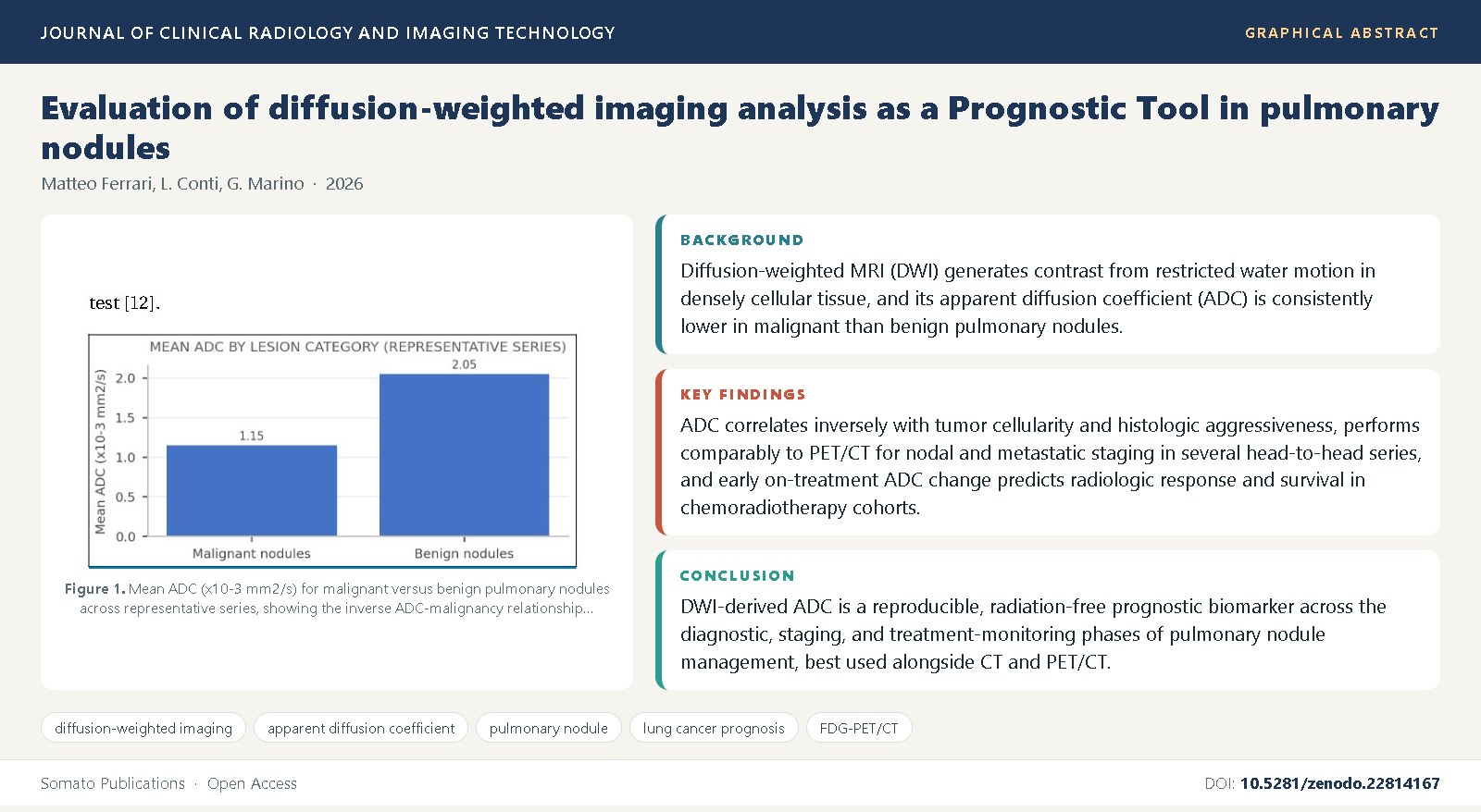
Background: Diffusion-weighted MRI (DWI) generates contrast from restricted water motion in densely cellular tissue, and its apparent diffusion coefficient (ADC) is consistently lower in malignant than benign pulmonary nodules.
Objective: This review evaluates DWI and ADC quantification as a prognostic tool in pulmonary nodules, covering nodal and distant staging, treatment-response prediction, survival correlation, and histogram and intravoxel incoherent motion refinements.
Methods: We synthesized diagnostic accuracy studies, prospective staging comparisons, treatment-response monitoring series, and meta-analyses addressing DWI, ADC, and IVIM parameters in pulmonary nodules and non-small cell lung cancer, benchmarked against PET/CT and CT.
Findings: ADC correlates inversely with tumor cellularity and histologic aggressiveness, performs comparably to PET/CT for nodal and metastatic staging in several head-to-head series, and early on-treatment ADC change predicts radiologic response and survival in chemoradiotherapy cohorts; histogram and radiomic refinements of mean ADC further improve discrimination.
Conclusions: DWI-derived ADC is a reproducible, radiation-free prognostic biomarker across the diagnostic, staging, and treatment-monitoring phases of pulmonary nodule management, best used alongside CT and PET/CT, with acquisition standardization remaining the principal barrier to routine prognostic adoption.
References
Le Bihan, D., Breton, E., Lallemand, D., Grenier, P., Cabanis, E., Laval-Jeantet, M. (1986) MR imaging of intravoxel incoherent motions: application to diffusion and perfusion in neurologic disorders. Radiology, 161(2): 401-407.
Koh, D.M., Collins, D.J. (2007) Diffusion-weighted MRI in the body: applications and challenges in oncology. American Journal of Roentgenology, 188(6): 1622-1635.
Padhani, A.R., Liu, G., Koh, D.M., Chenevert, T.L., Thoeny, H.C., Takahara, T., et al. (2009) Diffusion-weighted magnetic resonance imaging as a cancer biomarker: consensus and recommendations. Neoplasia, 11(2): 102-125.
Mori, T., Nomori, H., Ikeda, K., Kawanaka, K., Shiraishi, S., Katahira, K., et al. (2008) Diffusion-weighted magnetic resonance imaging for diagnosing malignant pulmonary nodules/masses: comparison with positron emission tomography. Journal of Thoracic Oncology, 3(4): 358-364.
Matoba, M., Tonami, H., Kondou, T., Yokota, H., Higashi, K., Toga, H., et al. (2007) Lung carcinoma: diffusion-weighted MR imaging--preliminary evaluation with apparent diffusion coefficient. Radiology, 243(2): 570-577.
Regier, M., Derlin, T., Schwarz, D., Laqmani, A., Henes, F.O., Groth, M., et al. (2011) Diffusion weighted MRI and 18F-FDG PET/CT in non-small cell lung cancer (NSCLC): does the apparent diffusion coefficient (ADC) correlate with tracer uptake (SUV)? European Journal of Radiology, 77(2): 297-303.
Liu, H., Liu, Y., Yu, T., Ye, N. (2010) Usefulness of diffusion-weighted MR imaging in the evaluation of pulmonary lesions. European Radiology, 20(4): 807-815.
Uto, T., Takehara, Y., Nakamura, Y., Naito, T., Hasegawa, M., Kamishima, T., et al. (2009) Higher sensitivity and specificity for diffusion-weighted imaging of malignant lung lesions without apparent diffusion coefficient quantification. Radiology, 252(1): 247-254.
Kim, H.J., Lee, K.Y., Kang, W.J., Kim, H.J., Choi, B.W. (2008) Solitary pulmonary nodules: correlation of apparent diffusion coefficients with tumor cellularity. AJR American Journal of Roentgenology, 191(6): 1701-1708.
Satoh, S., Kitazume, Y., Ohdama, S., Kimula, Y., Taura, S., Endo, Y. (2008) Can malignant and benign pulmonary nodules be differentiated with diffusion-weighted MRI? American Journal of Roentgenology, 191(2): 464-470.
Gumustas, S., Inan, N., Akansel, G., Ciftci, E., Demirci, A., Ozkan, F., et al. (2013) Differentiation of malignant and benign lung lesions with diffusion-weighted MR imaging. Radiology and Oncology, 47(2): 125-129.
Tanaka, R., Horikoshi, H., Nakazato, Y., Nagaki, N., Fukuda, T., Katsura, H., et al. (2009) Magnetic resonance imaging in diagnosis of solitary pulmonary nodules: comparison with computed tomography and positron emission tomography. Journal of Thoracic Imaging, 24(2): 90-95.
Razek, A.A., Fathy, A., Gawad, T.A. (2011) Correlation of apparent diffusion coefficient at 3T with prognostic parameters of lung cancer. AJR American Journal of Roentgenology, 196(4): W629-W636.
Chen, L., Zhang, J., Chen, Y., Wang, W., Zhou, X., Yan, X., et al. (2014) Relationship between apparent diffusion coefficient and tumour cellularity in lung cancer. PLoS ONE, 9(6): e99865.
Shen, G., Jia, Z., Deng, H. (2016) Apparent diffusion coefficient values of diffusion-weighted imaging for distinguishing focal pulmonary lesions and characterizing the subtype of lung cancer: a meta-analysis. European Radiology, 26(2): 556-566.
Nomori, H., Mori, T., Ikeda, K., Kawanaka, K., Shiraishi, S., Katahira, K., et al. (2008) Diffusion-weighted magnetic resonance imaging can be used in place of positron emission tomography for N staging of non-small cell lung cancer with fewer false-positive results. Journal of Thoracic and Cardiovascular Surgery, 135(4): 816-822.
Ohno, Y., Koyama, H., Yoshikawa, T., Matsumoto, K., Aoyama, N., Onishi, Y., et al. (2011) N stage disease in patients with non-small cell lung cancer: efficacy of quantitative and qualitative assessment with STIR turbo spin-echo imaging, diffusion-weighted MR imaging, and fluorodeoxyglucose PET/CT. Radiology, 261(2): 605-615.
Ohno, Y., Koyama, H., Onishi, Y., Takenaka, D., Nogami, M., Yoshikawa, T., et al. (2008) Non-small cell lung cancer: whole-body MR examination for M-stage assessment--utility for whole-body diffusion-weighted imaging compared with integrated FDG PET/CT. Radiology, 248(2): 643-654.
Wu, L.M., Xu, J.R., Gu, H.Y., Hua, J., Zhu, J., Chen, J., et al. (2012) Preoperative mediastinal and hilar nodal staging with diffusion-weighted magnetic resonance imaging and fluorodeoxyglucose positron emission tomography/computed tomography in patients with non-small-cell lung cancer: which is better? Journal of Surgical Research, 178(1): 304-314.
Usuda, K., Zhao, X.T., Sagawa, M., Matoba, M., Kuginuki, Y., Ueda, Y., et al. (2011) Diffusion-weighted imaging is superior to PET in the detection and nodal assessment of lung cancers. Annals of Thoracic Surgery, 91(6): 1689-1695.
Yang, R.M., Li, L., Wei, X.H., Guo, Y.M., Huang, Y.H., Lai, L.S., et al. (2014) Differentiation of central lung cancer from atelectasis: comparison of diffusion-weighted MRI with PET/CT. PLoS ONE, 8(4): e60279.
Ohno, Y., Koyama, H., Yoshikawa, T., Nishio, M., Aoyama, N., Onishi, Y., et al. (2012) Diffusion-weighted MRI versus 18F-FDG PET/CT: performance as predictors of tumor treatment response and patient survival in patients with non-small cell lung cancer receiving chemoradiotherapy. American Journal of Roentgenology, 198(1): 75-82.
Yabuuchi, H., Hatakenaka, M., Takayama, K., Matsuo, Y., Sunami, S., Kamitani, T., et al. (2011) Non-small cell lung cancer: detection of early response to chemotherapy by using contrast-enhanced dynamic and diffusion-weighted MR imaging. Radiology, 261(2): 598-604.
Zhang, M., Kono, M. (2019) Solitary pulmonary nodules: diffusion-weighted MR imaging and monitoring of response to chemotherapy. Cancer Imaging, 19(1): 1-9.
Luo, S., Yu, X.P., Yuan, X.R., Wang, X.M., Wu, Z.J., Chen, B., et al. (2013) Advanced non-small cell lung cancer: response evaluation with diffusion-weighted MRI. European Journal of Radiology, 82(11): 1806-1811.
Coolen, J., De Keyzer, F., Nafteux, P., De Leyn, P., Vansteenkiste, J., Dooms, C., et al. (2012) Malignant pleural disease: diagnosis by using diffusion-weighted and dynamic contrast-enhanced MR imaging--initial experience. Radiology, 263(3): 884-892.
Deng, Y., Yang, X., Li, X., Chen, Q., Huang, C., Yang, M., et al. (2018) Histogram analysis of diffusion-weighted imaging for the differentiation of lung cancer and benign pulmonary lesions. Journal of Thoracic Disease, 10(4): 2189-2197.
Yuan, M., Zhang, Y.D., Pu, X.H., Zhong, Y., Li, H., Wu, J.F., et al. (2017) Comparison of a radiomic biomarker with volumetric analysis for decoding tumour phenotypes of lung adenocarcinoma with different disease-free survival. European Radiology, 27(11): 4857-4865.
Wang, L.L., Lin, J., Liu, K., Chen, C.Z., Liu, H.Y., Lv, P., et al. (2014) Intravoxel incoherent motion diffusion-weighted MR imaging in differentiation of lung cancer from obstructive lung consolidation: comparison and correlation with pharmacokinetic parameters from dynamic contrast-enhanced MR imaging. European Radiology, 24(8): 1914-1922.
Wang, S., Dougherty, L., Tsourkas, A., Yang, W., Schnall, M.D. (2011) Diffusion-weighted MRI in lung cancer at 3T: reproducibility of apparent diffusion coefficient measurement. American Journal of Roentgenology, 197(1): W96-W101.
Peerlings, J., Troost, E.G., Nelemans, P.J., Cook, S.A., de Jong, J.J., Franssen, F.M., et al. (2019) The diagnostic value of MR imaging in determining the malignancy of solitary pulmonary nodules: a systematic review and meta-analysis. Radiology, 290(1): 217-226.

Published
Issue
Section
License
Copyright (c) 2026 Matteo Ferrari, et al.

This work is licensed under a Creative Commons Attribution 4.0 International License.