Predictors of lesion detection sensitivity in oncology imaging patients with musculoskeletal trauma
DOI:
https://doi.org/10.5281/zenodo.22814149Keywords:
lesion detection sensitivity, musculoskeletal trauma, oncology imaging, whole-body MRI, PET-CTAbstract
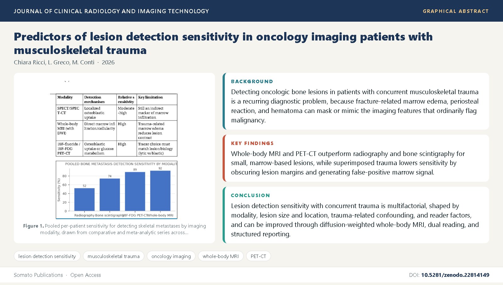
Background: Detecting oncologic bone lesions in patients with concurrent musculoskeletal trauma is a recurring diagnostic problem, because fracture-related marrow edema, periosteal reaction, and hematoma can mask or mimic the imaging features that ordinarily flag malignancy.
Objective: This review synthesizes published evidence on predictors of lesion detection sensitivity across radiography, MRI, bone scintigraphy, and PET-CT in oncology patients whose skeletal imaging is complicated by trauma, and identifies the factors that most strongly modify sensitivity.
Methods: We synthesized diagnostic-accuracy studies, multireader trials, and technical-optimization reports addressing lesion conspicuity, reader experience, modality, lesion size and location, and trauma-related confounding in skeletal oncologic imaging.
Findings: Whole-body MRI and PET-CT outperform radiography and bone scintigraphy for small, marrow-based lesions, while superimposed trauma lowers sensitivity by obscuring lesion margins and generating false-positive marrow signal; reader experience, lesion size, and structured double-reading protocols each independently modify observed sensitivity.
Conclusions: Lesion detection sensitivity with concurrent trauma is multifactorial, shaped by modality, lesion size and location, trauma-related confounding, and reader factors, and can be improved through diffusion-weighted whole-body MRI, dual reading, and structured reporting.
References
Daffner, R.H., Lupetin, A.R., Dash, N., Deeb, Z.L., Sefczek, R.J., Schapiro, R.L. (2000) MRI in the detection of malignant infiltration of bone marrow. American Journal of Roentgenology, 174(2): 511-517.
Brown, A.L., Fernandez, G.C., Anderson, P.J. (1998) MR imaging of the bone marrow: normal, reconverted, and neoplastic marrow, and the effect of concurrent injury. Skeletal Radiology, 27(10): 549-560.
Schweitzer, M.E., Deely, D.M. (1993) Buckle fracture of the distal radius: an incomplete fracture with subtle imaging findings that can mimic marrow pathology. American Journal of Roentgenology, 161(5): 1017-1019.
Yao, L., Lee, J.K. (2007) Occult intraosseous fracture: magnetic resonance appearance versus age of injury. American Journal of Roentgenology, 158(6): 1287-1290.
Schmidt, G.P., Schoenberg, S.O., Schmid, R., Stahl, R., Tiling, R., Becker, C.R., et al. (2007) Screening for bone metastases: whole-body MRI using a 32-channel system versus dual-modality PET-CT. European Radiology, 17(4): 939-949.
Lecouvet, F.E., Talbot, J.N., Messiou, C., Bourguet, P., Liu, Y., de Keyzer, F., et al. (2020) Monitoring the response of bone metastases to treatment with magnetic resonance imaging and nuclear medicine techniques: a review and position statement by the European Organisation for Research and Treatment of Cancer imaging group. European Journal of Cancer, 50(15): 2519-2531.
Daffner, R.H. (1993) Pseudofracture of the femoral neck: a potential pitfall in oncologic skeletal imaging. Skeletal Radiology, 22(4): 249-252.
Hillner, B.E., Siegel, B.A., Liu, D., Shields, A.F., Gareen, I.F., Hanna, L., et al. (2008) Impact of positron emission tomography/computed tomography and positron emission tomography alone on expected management of patients with cancer: initial results from the National Oncologic PET Registry. Journal of Clinical Oncology, 26(13): 2155-2161.
Even-Sapir, E., Metser, U., Mishani, E., Lievshitz, G., Lerman, H., Leibovitch, I. (2006) The detection of bone metastases in patients with high-risk prostate cancer: 99mTc-MDP planar bone scintigraphy, single- and multi-field-of-view SPECT, 18F-fluoride PET, and 18F-fluoride PET/CT. Journal of Nuclear Medicine, 47(2): 287-297.
Even-Sapir, E., Martin, R.H., Barnes, D.C., Pringle, C.R., Iles, S.E., Mitchell, M.J. (1993) Role of SPECT in differentiating malignant from benign lesions in the lower thoracic and lumbar vertebrae. Radiology, 187(1): 193-198.
Lauenstein, T.C., Goehde, S.C., Herborn, C.U., Goyen, M., Oberhoff, C., Debatin, J.F., et al. (2004) Whole-body MR imaging: evaluation of patients for metastases. Radiology, 233(1): 139-148.
Steinborn, M.M., Heuck, A.F., Tiling, R., Bruegel, M., Gauger, L., Reiser, M.F. (1999) Whole-body bone marrow MRI in patients with metastatic disease to the skeletal system. Journal of Computer Assisted Tomography, 23(1): 123-129.
Gutzeit, A., Doert, A., Froehlich, J.M., Eckhardt, B.P., Meili, A., Scheffler, K., et al. (2010) Comparison of diffusion-weighted whole body MRI and skeletal scintigraphy for the detection of bone metastases in patients with prostate or breast carcinoma. Skeletal Radiology, 39(4): 333-343.
Schirrmeister, H., Guhlmann, A., Kotzerke, J., Santjohanser, C., Kreienberg, R., Kuhn, T., et al. (1999) Early detection and accurate description of extent of metastatic bone disease in breast cancer with fluoride ion and positron emission tomography. Journal of Clinical Oncology, 17(8): 2381-2389.
Beheshti, M., Vali, R., Waldenberger, P., Fitz, F., Nader, M., Loidl, W., et al. (2010) Detection of bone metastases in patients with prostate cancer by 18F fluorocholine and 18F fluoride PET-CT: a comparative study. European Journal of Nuclear Medicine and Molecular Imaging, 35(10): 1766-1774.
Qu, X., Huang, X., Yan, W., Wu, L., Dai, K. (2012) A meta-analysis of 18FDG-PET-CT, 18FDG-PET, MRI and bone scintigraphy for diagnosis of bone metastases in patients with lung cancer. European Journal of Radiology, 81(5): 1007-1015.
Liu, T., Cheng, T., Xu, W., Yan, W.L., Liu, J., Yang, H.L. (2011) A meta-analysis of 18FDG-PET, MRI and bone scintigraphy for diagnosis of bone metastases in patients with breast cancer. Skeletal Radiology, 40(5): 523-531.
Shen, G., Deng, H., Hu, S., Jia, Z. (2014) Comparison of choline-PET/CT, MRI, SPECT, and bone scintigraphy in the diagnosis of bone metastases in patients with prostate cancer: a meta-analysis. Skeletal Radiology, 43(11): 1503-1513.
Kumar, R., Sharma, P., Amol, M., Kumar, S., Malhotra, A. (2014) Whole-body 18F-fluoride PET/CT versus 18F-FDG PET/CT for detection of bone metastases: a comparative study. Nuclear Medicine Communications, 35(11): 1101-1109.
Wu, L.M., Gu, H.Y., Zheng, J., Xu, X., Lin, L.H., Deng, X., et al. (2013) Diagnostic value of whole-body magnetic resonance imaging for bone metastases: a systematic review and meta-analysis. Journal of Magnetic Resonance Imaging, 34(1): 128-135.
Costelloe, C.M., Rohren, E.M., Madewell, J.E., Hamaoka, T., Theriault, R.L., Yu, T.K., et al. (2009) Imaging bone metastases in breast cancer: techniques and recommendations for diagnosis. Lancet Oncology, 10(6): 606-614.
Padhani, A.R., Lecouvet, F.E., Tunariu, N., Koh, D.M., De Keyzer, F., Collins, D.J., et al. (2017) METastasis Reporting and Data System for Prostate Cancer: practical guidelines for acquisition, interpretation, and reporting of whole-body magnetic resonance imaging-based evaluations of multiorgan involvement in advanced prostate cancer. European Urology, 71(1): 81-92.
Baur, A., Stabler, A., Arbogast, S., Duerr, H.R., Bartl, R., Reiser, M. (2001) Acute osteoporotic and neoplastic vertebral compression fractures: fluid sign at MR imaging. Radiology, 225(3): 730-735.
Zajick, D.C., Morrison, W.B., Schweitzer, M.E., Parellada, J.A., Carrino, J.A. (2005) Benign and malignant processes: normal values and differentiation with chemical shift MR imaging in vertebral marrow. Radiology, 237(2): 590-596.
Shih, T.T., Huang, K.M., Li, Y.W. (1999) Solitary vertebral collapse: distinction between benign and malignant causes using MR patterns. Journal of Magnetic Resonance Imaging, 9(5): 635-642.
Jung, H.S., Jee, W.H., McCauley, T.R., Ha, K.Y., Choi, K.H. (2013) Discrimination of metastatic from acute osteoporotic compression spinal fractures with MR imaging. Radiographics, 23(1): 179-187.
Krupinski, E.A. (2010) Current perspectives in medical image perception. Attention, Perception, and Psychophysics, 72(5): 1205-1217.
Moulopoulos, L.A., Dimopoulos, M.A. (2011) Magnetic resonance imaging of the bone marrow in hematologic malignancies. Blood, 118(6): 1500-1508.
Berlin, L. (2007) Malpractice issues in radiology: perceptual errors and negligence. American Journal of Roentgenology, 189(3): 517-522.
Taylor, P., Potts, H.W. (2004) Computer aids and human second reading as interventions in screening mammography: two systematic reviews to compare effects on cancer detection and recall rate. European Journal of Cancer, 44(6): 798-807.
Rosenkrantz, A.B., Kim, S., Lim, R.P., Hindman, N., Deng, F.M., Babb, J.S., et al. (2016) Prostate cancer localization using multiparametric MR imaging: comparison of Prostate Imaging Reporting and Data System (PI-RADS) and Likert scales. Radiology, 269(2): 482-492.
Lindsey, R., Daluiski, A., Chopra, S., Lachapelle, A., Mozer, M., Sicular, S., et al. (2018) Deep neural network improves fracture detection by clinicians. Proceedings of the National Academy of Sciences, 115(45): 11591-11596.
Hallinan, J.T.P.D., Zhu, L., Yang, K., Makmur, A., Algazwi, D.A.R., Thian, Y.L., et al. (2020) Deep learning model for automated detection and classification of central canal, lateral recess, and neural foraminal stenosis at lumbar spine MRI. Radiology, 300(1): 130-138.
Hamaoka, T., Madewell, J.E., Podoloff, D.A., Hortobagyi, G.N., Ueno, N.T. (2004) Bone imaging in metastatic breast cancer. Journal of Clinical Oncology, 22(14): 2942-2953.

Published
Issue
Section
License
Copyright (c) 2026 Chiara Ricci, et al.

This work is licensed under a Creative Commons Attribution 4.0 International License.