Doppler ultrasound assessment in the Management of endometriosis among pregnant women in low-resource settings
DOI:
https://doi.org/10.5281/zenodo.22813823Keywords:
Doppler ultrasound, endometriosis, pregnancy, low-resource settings, obstetric surveillanceAbstract
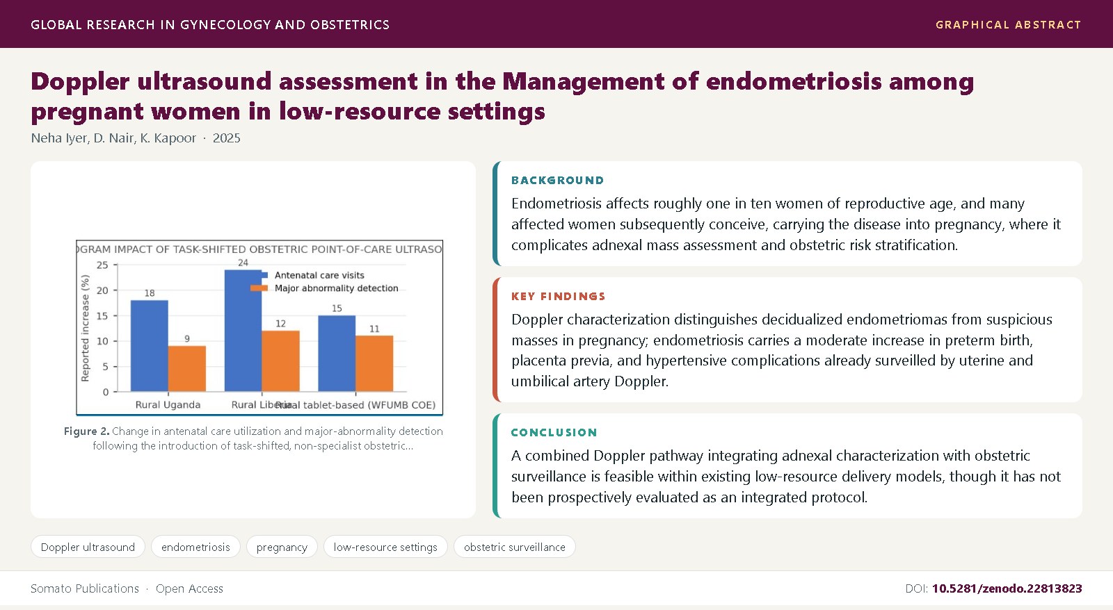
Background: Endometriosis affects roughly one in ten women of reproductive age, and many affected women subsequently conceive, carrying the disease into pregnancy, where it complicates adnexal mass assessment and obstetric risk stratification.
Objective: This review synthesizes evidence on Doppler ultrasound assessment of endometriosis in pregnant women, covering decidualized-endometrioma characterization, obstetric Doppler surveillance for endometriosis-associated complications, and feasibility in low-resource settings.
Methods: We conducted a narrative synthesis of consensus terminology statements, diagnostic-accuracy reviews, cohort studies of obstetric outcomes, and implementation studies of point-of-care obstetric ultrasound in low- and middle-income countries.
Findings: Doppler characterization distinguishes decidualized endometriomas from suspicious masses in pregnancy; endometriosis carries a moderate increase in preterm birth, placenta previa, and hypertensive complications already surveilled by uterine and umbilical artery Doppler; and task-shifted point-of-care ultrasound programs show these techniques can be delivered by non-specialist operators in resource-limited settings.
Conclusions: A combined Doppler pathway integrating adnexal characterization with obstetric surveillance is feasible within existing low-resource delivery models, though it has not been prospectively evaluated as an integrated protocol and remains constrained by supervision gaps documented across implementation programs.
References
Zondervan, K.T., Becker, C.M., Missmer, S.A. (2020) Endometriosis. New England Journal of Medicine, 382(13): 1244-1256. DOI: 10.1056/NEJMra1810764
Giudice, L.C. (2010) Clinical practice. Endometriosis. New England Journal of Medicine, 362(25): 2389-2398. DOI: 10.1056/NEJMcp1000274
Missmer, S.A., Hankinson, S.E., Spiegelman, D., Barbieri, R.L., Marshall, L.M., Hunter, D.J. (2004) Incidence of laparoscopically confirmed endometriosis by demographic, anthropometric, and lifestyle factors. American Journal of Epidemiology, 160(8): 784-796. DOI: 10.1093/aje/kwh275
Farquhar, C. (2007) Endometriosis. BMJ, 334(7587): 249-253. DOI: 10.1136/bmj.39073.736829.BE
Vercellini, P., Vigano, P., Somigliana, E., Fedele, L. (2014) Endometriosis: pathogenesis and treatment. Nature Reviews Endocrinology, 10(5): 261-275. DOI: 10.1038/nrendo.2013.255
Guerriero, S., Condous, G., van den Bosch, T., Valentin, L., Leone, F.P., Van Schoubroeck, D., et al. (2016) Systematic approach to sonographic evaluation of the pelvis in women with suspected endometriosis, including terms, definitions and measurements: a consensus opinion from the International Deep Endometriosis Analysis (IDEA) group. Ultrasound in Obstetrics and Gynecology, 48(3): 318-332. DOI: 10.1002/uog.15955
Exacoustos, C., Manganaro, L., Zupi, E. (2014) Imaging for the evaluation of endometriosis and adenomyosis. Best Practice and Research Clinical Obstetrics and Gynaecology, 28(5): 655-681. DOI: 10.1016/j.bpobgyn.2014.04.010
Chapron, C., Fauconnier, A., Vieira, M., Barakat, H., Dousset, B., Pansini, V., et al. (2003) Anatomical distribution of deep infiltrating endometriosis: surgical implications and proposition for a classification. Human Reproduction, 18(1): 157-161. DOI: 10.1093/humrep/deg009
Bazot, M., Darai, E. (2017) Diagnosis of deep endometriosis: clinical examination, ultrasonography, magnetic resonance imaging, and other techniques. Fertility and Sterility, 108(6): 886-894. DOI: 10.1016/j.fertnstert.2017.10.026
Hudelist, G., English, J., Thomas, A.E., Tinelli, A., Singer, C.F., Keckstein, J. (2011) Diagnostic accuracy of transvaginal ultrasound for non-invasive diagnosis of bowel endometriosis: systematic review and meta-analysis. Ultrasound in Obstetrics and Gynecology, 37(3): 257-263. DOI: 10.1002/uog.8858
Nisenblat, V., Bossuyt, P.M., Farquhar, C., Johnson, N., Hull, M.L. (2016) Imaging modalities for the non-invasive diagnosis of endometriosis. Cochrane Database of Systematic Reviews, 2: CD009591. DOI: 10.1002/14651858.CD009591.pub2
Leone, F.P., Timmerman, D., Bourne, T., Valentin, L., Epstein, E., Goldstein, S.R., et al. (2010) Terms, definitions and measurements to describe the sonographic features of adnexal tumors: a consensus opinion from the International Ovarian Tumor Analysis (IOTA) group. Ultrasound in Obstetrics and Gynecology, 35(1): 103-112. DOI: 10.1002/uog.7487
Van Calster, B., Van Hoorde, K., Valentin, L., Testa, A.C., Fischerova, D., Van Holsbeke, C., et al. (2014) Evaluating the risk of ovarian cancer before surgery using the ADNEX model to differentiate between benign, borderline, early and advanced stage invasive, and secondary metastatic tumours: prospective multicentre diagnostic study. BMJ, 349: g5920. DOI: 10.1136/bmj.g5920
Mascilini, F., Moruzzi, C., Giansiracusa, C., Guastafierro, F., Savelli, L., De Meis, L., et al. (2014) Imaging in gynecological disease (10): clinical and ultrasound characteristics of decidualized endometriomas surgically removed during pregnancy. Ultrasound in Obstetrics and Gynecology, 44(3): 354-360. DOI: 10.1002/uog.13323
Leeners, B., Damaso, F., Ochsenbein-Kolble, N., Farquhar, C. (2018) The effect of pregnancy on endometriosis-facts or fiction? Human Reproduction Update, 24(3): 290-299. DOI: 10.1093/humupd/dmy004
Lalani, S., Choudhry, A.J., Firth, B., Bacal, V., Walker, M., Wen, S.W., et al. (2018) Endometriosis and adverse maternal, fetal and neonatal outcomes, a systematic review and meta-analysis. Human Reproduction, 33(10): 1854-1865. DOI: 10.1093/humrep/dey269
Saraswat, L., Ayansina, D.T., Cooper, K.G., Bhattacharya, S., Miligkos, D., Bhattacharya, S. (2017) Pregnancy outcomes in women with endometriosis: a national record linkage study. BJOG, 124(3): 444-452. DOI: 10.1111/1471-0528.13920
Glavind, M.T., Forman, A., Arendt, L.H., Nielsen, K., Henriksen, T.B. (2017) Endometriosis and pregnancy complications: a Danish cohort study. Fertility and Sterility, 107(1): 160-166. DOI: 10.1016/j.fertnstert.2016.09.020
Harada, T., Taniguchi, F., Amano, H., Kurozawa, Y., Ideno, Y., Hayashi, K., et al. (2019) Adverse obstetric outcomes for women with endometriosis and adenomyosis: a large cohort of the Japan Environment and Children's Study. PLoS One, 14(8): e0220256. DOI: 10.1371/journal.pone.0220256
Swanson, J.O., Kawooya, M.G., Swanson, D.L., Hippe, D.S., Dungu-Matovu, P., Nathan, R. (2014) The diagnostic impact of limited, screening obstetric ultrasound when performed by midwives in rural Uganda. Journal of Perinatology, 34(7): 508-512. DOI: 10.1038/jp.2014.54
Vinayak, S., Sande, J., Nisenbaum, H., Nolsoe, C.P. (2017) Training midwives to perform basic obstetric point-of-care ultrasound in rural areas using a tablet platform and mobile phone transmission technology - a WFUMB COE project. Ultrasound in Medicine and Biology, 43(10): 2125-2132. DOI: 10.1016/j.ultrasmedbio.2017.05.024
Ross, A.B., DeStigter, K.K., Rielly, M., Souza, S., Morey, G.E., Nelson, M., et al. (2013) A low-cost ultrasound program leads to increased antenatal care utilization in rural Liberia. PLoS One, 8(10): e78450. DOI: 10.1371/journal.pone.0078450
Kim, E.T., Singh, K., Moran, A., Armbruster, D., Kozuki, N. (2018) Obstetric ultrasound use in low and middle income countries: a narrative review. Reproductive Health, 15(1): 129. DOI: 10.1186/s12978-018-0571-y
Exacoustos, C., Morosetti, G., Conway, F., Camilli, S., Martinez, D., Alesi, M., et al. (2018) New sonographic classification of adenomyosis to predict severity of dysmenorrhea and menorrhagia. Journal of Minimally Invasive Gynecology, 25(7): 1259-1266. DOI: 10.1016/j.jmig.2018.03.023
Reid, S., Lu, C., Casikar, I., Reid, G., Abbott, J., Cario, G., et al. (2013) Prediction of pouch of Douglas obliteration in women with suspected endometriosis using a new real-time dynamic transvaginal ultrasound technique: the sliding sign. Ultrasound in Obstetrics and Gynecology, 41(6): 685-691. DOI: 10.1002/uog.12305
Campbell, S., Griffin, D.R., Pearce, J.M., Diaz-Recasens, J., Cohen-Overbeek, T.E., Willson, K., et al. (1983) New Doppler technique for assessing uteroplacental blood flow. Lancet, 1(8326): 675-677. DOI: 10.1016/S0140-6736(83)91970-0
Papageorghiou, A.T., Yu, C.K., Bindra, R., Pandis, G., Nicolaides, K.H. (2001) Multicenter screening for pre-eclampsia and fetal growth restriction by transvaginal uterine artery Doppler at 23 weeks of gestation. Ultrasound in Obstetrics and Gynecology, 18(5): 441-449. DOI: 10.1046/j.0960-7692.2001.00572.x
Cnossen, J.S., Morris, R.K., ter Riet, G., Mol, B.W., van der Post, J.A., Coomarasamy, A., et al. (2008) Use of uterine artery Doppler ultrasonography to predict pre-eclampsia and intrauterine growth restriction: a systematic review and bivariable meta-analysis. CMAJ, 178(6): 701-711. DOI: 10.1503/cmaj.070430
Alfirevic, Z., Stampalija, T., Gyte, G.M. (2013) Fetal and umbilical Doppler ultrasound in high-risk pregnancies. Cochrane Database of Systematic Reviews, 11: CD007529. DOI: 10.1002/14651858.CD007529.pub3
Alfirevic, Z., Neilson, J.P. (1995) Doppler ultrasonography in high-risk pregnancies: systematic review with meta-analysis. American Journal of Obstetrics and Gynecology, 172(5): 1379-1387. DOI: 10.1016/0002-9378(95)90466-2
World Health Organization (2016) WHO recommendations on antenatal care for a positive pregnancy experience. Geneva: World Health Organization.
Shah, S., Bellows, B.A., Adedipe, A.A., Totten, J.E., Backlund, B.H., Sajed, D. (2015) Perceived barriers in the use of ultrasound in developing countries. Critical Ultrasound Journal, 7: 11. DOI: 10.1186/s13089-015-0028-2
Becker, C.M., Bokor, A., Heikinheimo, O., Horne, A., Jansen, F., Kiesel, L., et al. (2022) ESHRE guideline: endometriosis. Human Reproduction Open, 2022(2): hoac009. DOI: 10.1093/hropen/hoac009

Published
Issue
Section
License
Copyright (c) 2025 Neha Iyer, et al.

This work is licensed under a Creative Commons Attribution 4.0 International License.