Evaluation of Doppler ultrasound assessment as a Prognostic Tool in gestational diabetes
DOI:
https://doi.org/10.5281/zenodo.22813741Keywords:
gestational diabetes, Doppler ultrasound, umbilical artery, cerebroplacental ratio, perinatal outcomeAbstract
Background: Glycemic control alone does not fully explain which GDM pregnancies progress to macrosomia or other adverse perinatal outcome, prompting interest in Doppler indices as adjunct prognostic markers.
Objective: To determine whether third-trimester umbilical artery, uterine artery and cerebroplacental ratio (CPR) Doppler indices independently predict adverse perinatal outcome in GDM, and whether a combined Doppler score adds discriminative value.
Methods: In a prospective cohort of singleton GDM pregnancies, umbilical, uterine and middle cerebral artery Doppler indices were obtained at 34-37 weeks and related to a composite outcome of macrosomia, non-reassuring caesarean delivery, low Apgar score, hypoglycemia or NICU admission by adjusted logistic regression.
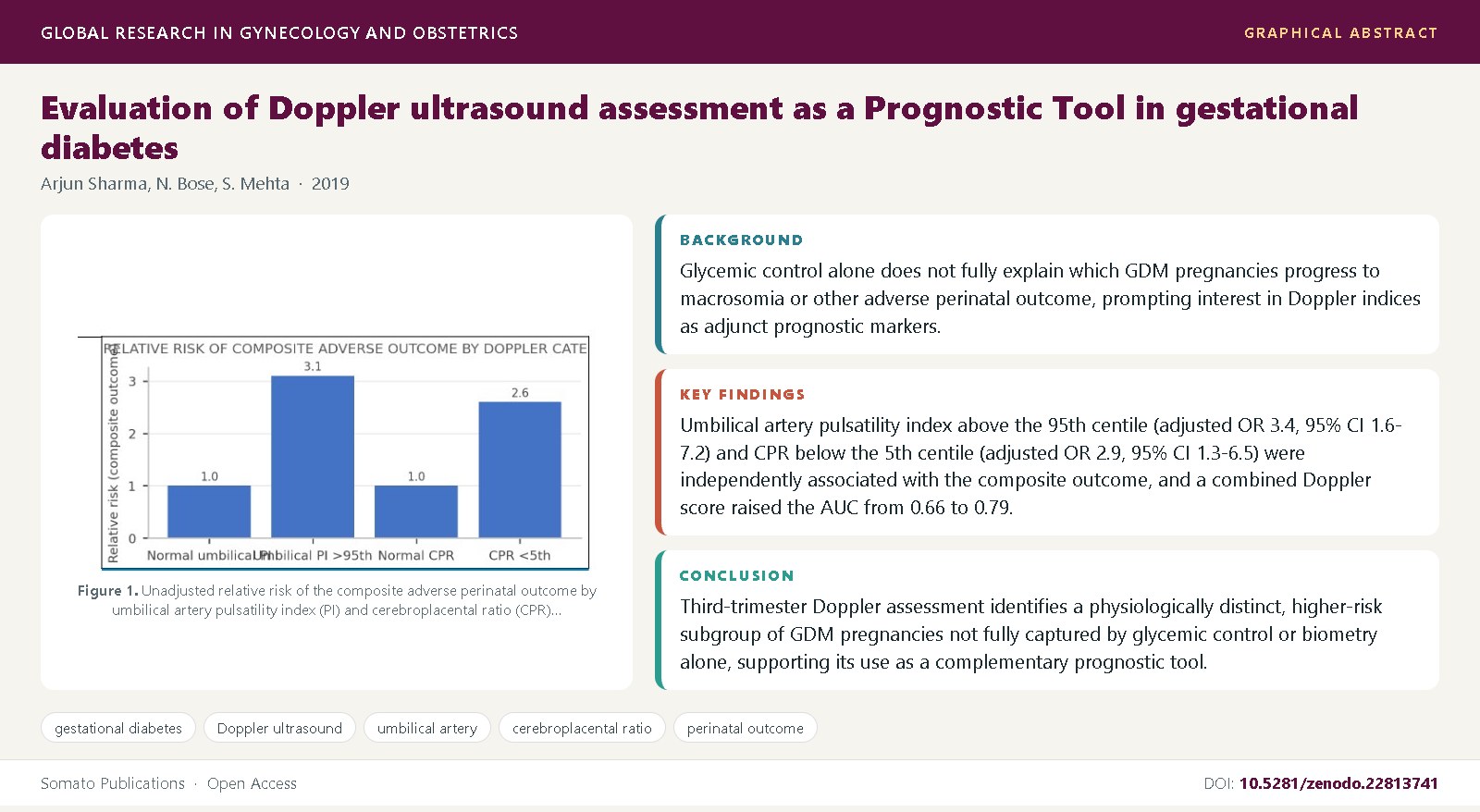
Results: Umbilical artery pulsatility index above the 95th centile (adjusted OR 3.4, 95% CI 1.6-7.2) and CPR below the 5th centile (adjusted OR 2.9, 95% CI 1.3-6.5) were independently associated with the composite outcome, and a combined Doppler score raised the AUC from 0.66 to 0.79.
Conclusions: Third-trimester Doppler assessment identifies a physiologically distinct, higher-risk subgroup of GDM pregnancies not fully captured by glycemic control or biometry alone, supporting its use as a complementary prognostic tool.
References
HAPO Study Cooperative Research Group, Metzger, B.E., Lowe, L.P., Dyer, A.R., Trimble, E.R., Chaovarindr, U., et al. (2008) Hyperglycemia and adverse pregnancy outcomes. New England Journal of Medicine, 358(19): 1991-2002. DOI: 10.1056/NEJMoa0707943
Crowther, C.A., Hiller, J.E., Moss, J.R., McPhee, A.J., Jeffries, W.S., Robinson, J.S. (2005) Effect of treatment of gestational diabetes mellitus on pregnancy outcomes. New England Journal of Medicine, 352(24): 2477-2486. DOI: 10.1056/NEJMoa042973
Landon, M.B., Spong, C.Y., Thom, E., Carpenter, M.W., Ramin, S.M., Casey, B., et al. (2009) A multicenter, randomized trial of treatment for mild gestational diabetes. New England Journal of Medicine, 361(14): 1339-1348. DOI: 10.1056/NEJMoa0902430
Langer, O., Yogev, Y., Most, O., Xenakis, E.M. (2005) Gestational diabetes: the consequences of not treating. American Journal of Obstetrics and Gynecology, 192(4): 989-997. DOI: 10.1016/j.ajog.2004.11.039
Catalano, P.M., Hauguel-De Mouzon, S. (2011) Is it time to revisit the Pedersen hypothesis in the face of the obesity epidemic? American Journal of Obstetrics and Gynecology, 204(6): 479-487. DOI: 10.1016/j.ajog.2010.11.039
American College of Obstetricians and Gynecologists. (2018) ACOG Practice Bulletin No. 190: Gestational diabetes mellitus. Obstetrics and Gynecology, 131(2): e49-e64. DOI: 10.1097/AOG.0000000000002501
Campbell, S., Diaz-Recasens, J., Griffin, D.R., Cohen-Overbeek, T.E., Pearce, J.M., Willson, K., et al. (1983) New doppler technique for assessing uteroplacental blood flow. Lancet, 1(8326): 675-677. DOI: 10.1016/s0140-6736(83)91970-0
Trudinger, B.J., Giles, W.B., Cook, C.M. (1985) Uteroplacental blood flow velocity-time waveforms in normal and complicated pregnancy. British Journal of Obstetrics and Gynaecology, 92(1): 39-45. DOI: 10.1111/j.1471-0528.1985.tb01046.x
Gudmundsson, S., Marsal, K. (1988) Umbilical and uteroplacental blood flow velocity waveforms in pregnancies with fetal growth retardation. European Journal of Obstetrics and Gynecology and Reproductive Biology, 27(3): 187-196. DOI: 10.1016/0028-2243(88)90128-1
Karsdorp, V.H., van Vugt, J.M., van Geijn, H.P., Kostense, P.J., Arduini, D., Montenegro, N., et al. (1994) Clinical significance of absent or reversed end diastolic velocity waveforms in umbilical artery. Lancet, 344(8938): 1664-1668. DOI: 10.1016/s0140-6736(94)90457-x
Baschat, A.A., Gembruch, U., Reiss, I., Gortner, L., Weiner, C.P., Harman, C.R. (2000) Relationship between arterial and venous Doppler and perinatal outcome in fetal growth restriction. Ultrasound in Obstetrics and Gynecology, 16(5): 407-413. DOI: 10.1046/j.1469-0705.2000.00284.x
Hershkovitz, R., Kingdom, J.C., Geary, M., Rodeck, C.H. (2000) Fetal cerebral blood flow redistribution in late gestation: identification of compromise in small fetuses with normal umbilical artery Doppler. Ultrasound in Obstetrics and Gynecology, 15(3): 209-212. DOI: 10.1046/j.1469-0705.2000.00079.x
Turan, O.M., Turan, S., Gungor, S., Berg, C., Moyano, D., Gembruch, U., et al. (2008) Progression of Doppler abnormalities in intrauterine growth restriction. Ultrasound in Obstetrics and Gynecology, 32(2): 160-167. DOI: 10.1002/uog.5386
Morales-Rosello, J., Khalil, A., Morlando, M., Papageorghiou, A., Bhide, A., Thilaganathan, B. (2014) Changes in fetal Doppler indices as a marker of failure to reach growth potential at term. Ultrasound in Obstetrics and Gynecology, 43(3): 303-310. DOI: 10.1002/uog.13319
Figueras, F., Gratacos, E. (2014) Update on the diagnosis and classification of fetal growth restriction and proposal of a stage-based management protocol. Fetal Diagnosis and Therapy, 36(2): 86-98. DOI: 10.1159/000357592
Cnossen, J.S., Morris, R.K., ter Riet, G., Mol, B.W., van der Post, J.A., Coomarasamy, A., et al. (2008) Use of uterine artery Doppler ultrasonography to predict pre-eclampsia and intrauterine growth restriction: a systematic review and bibliometric analysis. Canadian Medical Association Journal, 178(6): 701-711. DOI: 10.1503/cmaj.070430
Alfirevic, Z., Neilson, J.P. (1995) Doppler ultrasonography in high-risk pregnancies: systematic review with meta-analysis. American Journal of Obstetrics and Gynecology, 172(5): 1379-1387. DOI: 10.1016/0002-9378(95)90466-2
Bhide, A., Acharya, G., Bilardo, C.M., Brezinka, C., Cafici, D., Hernandez-Andrade, E., et al. (2013) ISUOG practice guidelines: use of Doppler ultrasonography in obstetrics. Ultrasound in Obstetrics and Gynecology, 41(2): 233-239. DOI: 10.1002/uog.12371
Salomon, L.J., Alfirevic, Z., Da Silva Costa, F., Deter, R.L., Figueras, F., Ghi, T., et al. (2019) ISUOG practice guidelines: ultrasound assessment of fetal biometry and growth. Ultrasound in Obstetrics and Gynecology, 53(6): 715-723. DOI: 10.1002/uog.20272
Nylund, L., Lunell, N.O., Lewander, R., Persson, B., Sarby, B. (1982) Uteroplacental blood flow in diabetic pregnancy: measurements with indium 113m and a computer-linked gamma camera. American Journal of Obstetrics and Gynecology, 144(3): 298-302. DOI: 10.1016/0002-9378(82)90581-6
Salvesen, D.R., Higueras, M.T., Mansur, C.A., Freeman, J., Brudenell, J.M., Nicolaides, K.H. (1993) Placental and fetal Doppler velocimetry in pregnancies complicated by maternal diabetes mellitus. American Journal of Obstetrics and Gynecology, 168(2): 645-652. DOI: 10.1016/0002-9378(93)90511-z
Grunewald, C., Divon, M., Lunell, N.O. (1996) Doppler velocimetry in last trimester pregnancy complicated by insulin-dependent diabetes mellitus. Acta Obstetricia et Gynecologica Scandinavica, 75(9): 804-808. DOI: 10.3109/00016349609054707
Bracero, L.A., Schulman, H., Fleischer, A., Farmakides, G., Rochelson, B. (1986) Umbilical artery velocimetry in diabetes and pregnancy. Obstetrics and Gynecology, 68(5): 654-658.
Zimmermann, P., Kujansuu, E., Tuimala, R. (1995) Doppler flow velocimetry of the umbilical artery, uteroplacental arteries and fetal middle cerebral artery in pregnancies complicated by type I diabetes mellitus. Journal of Perinatal Medicine, 23(3): 215-223. DOI: 10.1515/jpme.1995.23.3.215
Leung, W.C., Lam, H., Lee, C.P., Lao, T.T. (2004) Doppler study of the umbilical and fetal middle cerebral arteries in women with gestational diabetes mellitus. Ultrasound in Obstetrics and Gynecology, 24(5): 534-537. DOI: 10.1002/uog.1740
Pietryga, M., Brazert, J., Wender-Ozegowska, E., Dubiel, M., Gudmundsson, S. (2006) Placental Doppler velocimetry in gestational diabetes mellitus. Journal of Perinatal Medicine, 34(2): 108-110. DOI: 10.1515/JPM.2006.019 disease. BMJ, 301(6761): 1111. DOI: 10.1136/bmj.301.6761.1111
Maruotti, G.M., Rizzo, G., Sirico, A., Sarno, L., Cirigliano, L., Arduini, D., et al. (2014) Are there any relationships between umbilical artery Doppler parameters and fetal growth in early-controlled diabetic pregnancies? Journal of Maternal-Fetal and Neonatal Medicine, 27(16): 1650-1654. DOI: 10.3109/14767058.2013.871704
American College of Obstetricians and Gynecologists. (2018) ACOG Practice Bulletin No. 201: Pregestational diabetes mellitus. Obstetrics and Gynecology, 132(6): e228-e248. DOI: 10.1097/AOG.0000000000002960
Maulik, D. (1989) Doppler ultrasound velocimetry for fetal surveillance. Clinical Obstetrics and Gynecology, 32(4): 671-689. DOI: 10.1097/00003081-198912000-00003
Metzger, B.E., Gabbe, S.G., Persson, B., Buchanan, T.A., Catalano, P.A., Damm, P., et al. (2010) International association of diabetes and pregnancy study groups recommendations on the diagnosis and classification of hyperglycemia in pregnancy. Diabetes Care, 33(3): 676-682. DOI: 10.2337/dc09-1848
Yoon, B.H., Lee, C.M., Kim, S.W. (1994) An abnormal umbilical artery waveform: a strong and independent predictor of adverse perinatal outcome in patients with preeclampsia. American Journal of Obstetrics and Gynecology, 171(3): 713-721. DOI: 10.1016/0002-9378(94)90084-1
Arduini, D., Rizzo, G. (1992) Prediction of fetal outcome in small for gestational age fetuses: comparison of Doppler measurements obtained from different fetal vessels. Journal of Perinatal Medicine, 20(1): 29-38. DOI: 10.1515/jpme.1992.20.1.29
Jansson, T., Ekstrand, Y., Bjorn, C., Wennergren, M., Powell, T.L. (2002) Alterations in the activity of placental amino acid transporters in pregnancies complicated by diabetes. Diabetes, 51(7): 2214-2219. DOI: 10.2337/diabetes.51.7.2214
Barker, D.J. (1990) The fetal and infant origins of adult

Published
Issue
Section
License
Copyright (c) 2019 Arjun Sharma, et al.

This work is licensed under a Creative Commons Attribution 4.0 International License.