
Comparative Analysis of corneal transplantation versus Standard Care in diabetic retinopathy
DOI:
https://doi.org/10.5281/zenodo.22811908Keywords:
corneal transplantation, diabetic retinopathy, keratoplasty, endothelial keratoplasty, anti-VEGF therapyAbstract
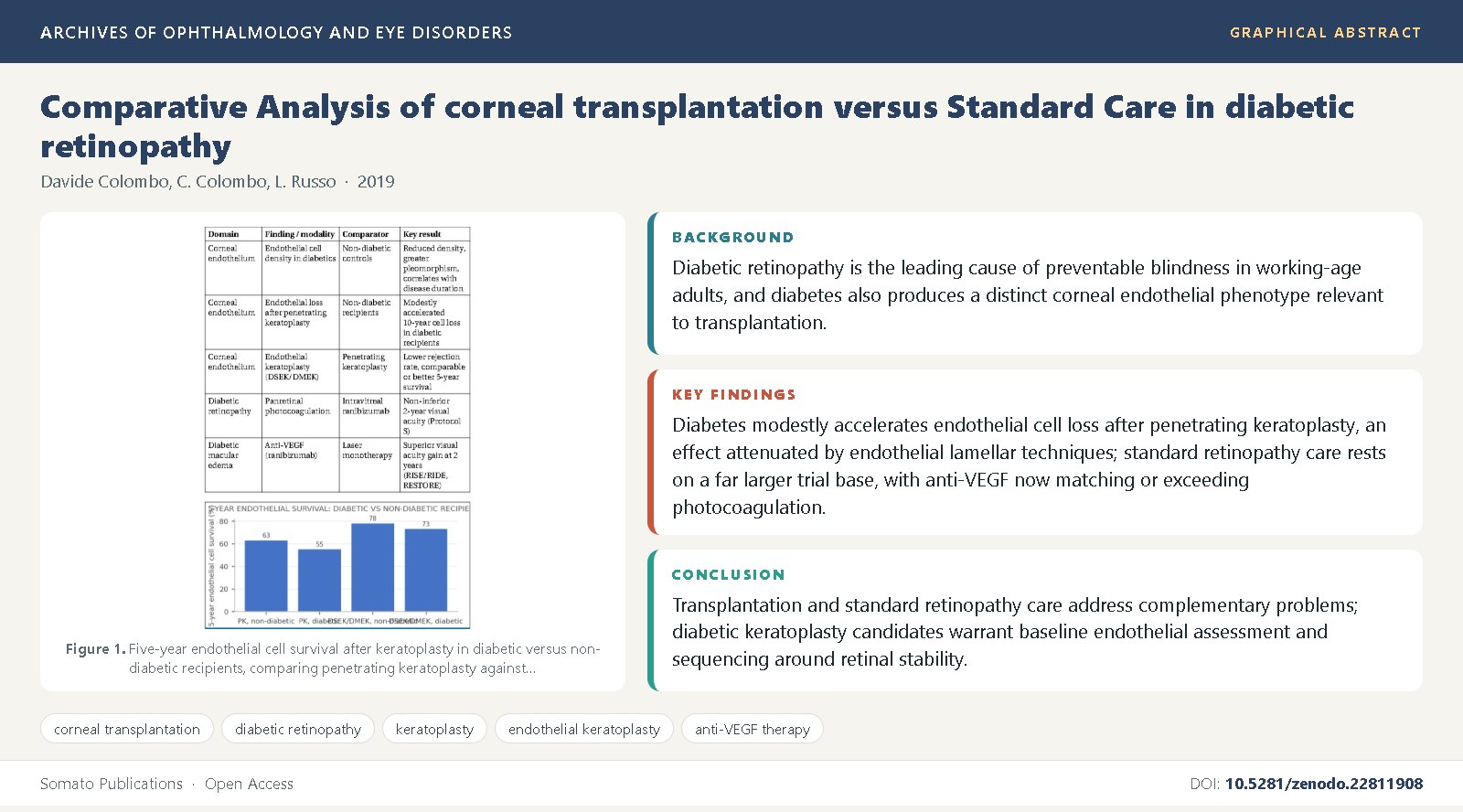
Background: Diabetic retinopathy is the leading cause of preventable blindness in working-age adults, and diabetes also produces a distinct corneal endothelial phenotype relevant to transplantation [6,7].
Objective: This review compares outcomes of corneal transplantation, including penetrating and endothelial keratoplasty, in diabetic recipients against standard, non-transplant care for diabetic retinopathy.
Methods: We synthesized keratoplasty outcome cohorts, eye-bank registry data, corneal endothelial physiology studies, and landmark diabetic retinopathy treatment trials from the PubMed-indexed ophthalmology literature.
Findings: Diabetes modestly accelerates endothelial cell loss after penetrating keratoplasty [21,23], an effect attenuated by endothelial lamellar techniques [26,25]; standard retinopathy care rests on a far larger trial base, with anti-VEGF now matching or exceeding photocoagulation [29,31].
Conclusions: Transplantation and standard retinopathy care address complementary problems; diabetic keratoplasty candidates warrant baseline endothelial assessment and sequencing around retinal stability [9,3].
References
Cheung, N., Mitchell, P., Wong, T.Y. (2010) Diabetic retinopathy. Lancet, 376(9735): 124-136. DOI: 10.1016/S0140-6736(09)62124-3
Yau, J.W., Rogers, S.L., Kawasaki, R., Lamoureux, E.L., Kowalski, J.W., Bek, T., et al. (2012) Global prevalence and major risk factors of diabetic retinopathy. Diabetes Care, 35(3): 556-564. DOI: 10.2337/dc11-1909
Wong, T.Y., Cheung, C.M., Larsen, M., Sharma, S., Simo, R. (2016) Diabetic retinopathy. Nature Reviews Disease Primers, 2: 16012. DOI: 10.1038/nrdp.2016.12
Aiello, L.P., Gardner, T.W., King, G.L., Blankenship, G., Cavallerano, J.D., Ferris, F.L., et al. (1998) Diabetic retinopathy. Diabetes Care, 21(1): 143-156. DOI: 10.2337/diacare.21.1.143
Klein, R., Klein, B.E., Moss, S.E., Cruickshanks, K.J. (1998) The Wisconsin Epidemiologic Study of Diabetic Retinopathy: XVII. The 14-year incidence and progression of diabetic retinopathy and associated risk factors in type 1 diabetes. Ophthalmology, 105(10): 1801-1815. DOI: 10.1016/S0161-6420(98)91020-X
Lee, J.S., Oum, B.S., Choi, H.Y., Lee, J.E., Cho, B.M. (2006) Differences in corneal thickness and corneal endothelium related to duration in diabetes. Eye, 20(3): 315-318. DOI: 10.1038/sj.eye.6701868
Sudhir, R.R., Raman, R., Sharma, T. (2012) Changes in the corneal endothelial cell density and morphology in patients with type 2 diabetes mellitus. Cornea, 31(10): 1119-1122. DOI: 10.1097/ICO.0b013e31823f8e26
Storr-Paulsen, A., Singh, A., Jeppesen, H., Norregaard, J.C., Thulesen, J. (2014) Corneal endothelial morphology and central thickness in patients with type II diabetes mellitus. Acta Ophthalmologica, 92(2): 158-160. DOI: 10.1111/aos.12064
Simo, R., Hernandez, C. (2014) Neurodegeneration in the diabetic eye: new insights and therapeutic perspectives. Trends in Endocrinology and Metabolism, 25(1): 23-33. DOI:
1016/j.tem.2013.09.005 10. Gorovoy, M.S. (2006) Descemet-stripping automated endothelial keratoplasty. Cornea, 25(8): 886-889. DOI: 10.1097/01.ico.0000214224.90743.01
Melles, G.R., Ong, T.S., Ververs, B., van der Wees, J. (2006) Descemet membrane endothelial keratoplasty (DMEK). Cornea, 25(8): 987-990. DOI: 10.1097/01.ico.0000248385.16896.34
Price, F.W., Price, M.O. (2009) Endothelial keratoplasty to restore clarity to a failed penetrating graft. Cornea, 28(1): 1-6. DOI: 10.1097/ICO.0b013e31817cd39c
Tan, D.T., Dart, J.K., Holland, E.J., Kinoshita, S. (2012) Corneal transplantation. Lancet, 379(9827): 1749-1761. DOI: 10.1016/S0140-6736(12)60437-1
Price, M.O., Mehta, J.S., Jurkunas, U.V., Price, F.W. (2018) Corneal endothelial dysfunction: evolving understanding and treatment options. Progress in Retinal and Eye Research, 65: 18-39. DOI: 10.1016/j.preteyeres.2018.03.001
Early Treatment Diabetic Retinopathy Study Research Group (1991) Early photocoagulation for diabetic retinopathy. ETDRS report number 9. Ophthalmology, 98(5 Suppl): 766-785.
Diabetes Control and Complications Trial Research Group (1993) The effect of intensive treatment of diabetes on the development and progression of long-term complications in insulin-dependent diabetes mellitus. New England Journal of Medicine, 329(14): 977-986. DOI: 10.1056/NEJM199309303291401
Diabetic Retinopathy Clinical Research Network (2015) Aflibercept, bevacizumab, or ranibizumab for diabetic macular edema. New England Journal of Medicine, 372(13): 1193-1203. DOI: 10.1056/NEJMoa1414264
Schultz, R.O., Van Horn, D.L., Peters, M.A., Klewin, K.M., Schutten, W.H. (1981) Diabetic keratopathy. Transactions of the American Ophthalmological Society, 79: 180-199.
Elner, S.G., Elner, V.M., Diaz-Rohena, R., Cameron, J.D., Stern, G.A., Tolentino, F.I. (1985) Corneal epithelial and stromal complications after diabetic vitrectomy. Ophthalmology, 92(9): 1198-1202.
Didenko, T.N., Smoleziaeva, G.L., Egorova, E.V. (1999) Corneal endothelial changes in diabetic patients undergoing cataract extraction. Vestnik Oftalmologii, 115(2): 21-23.
Lass, J.H., Benetz, B.A., Gal, R.L., Kollman, C., Raghinaru, D., Dontchev, M., et al. (2013) Donor age and factors related to endothelial cell loss 10 years after penetrating keratoplasty: Specular Microscopy Ancillary Study. Ophthalmology, 120(12): 2428-2435. DOI: 10.1016/j.ophtha.2013.08.044
Cornea Donor Study Investigator Group (2008) The effect of donor age on corneal transplantation outcome: results of the cornea donor study. Cornea, 27(5): 605-610. DOI: 10.1097/ICO.0b013e31816f0a19
Musch, D.C., Niziol, L.M., Stinnett, S.S., Kalayoglu, M. (2011) Contribution of diabetes to slow endothelial cell loss after penetrating keratoplasty. Cornea, 30(6): 685-689.
Bourne, W.M., Hodge, D.O., Nelson, L.R. (1994) Corneal endothelium five years after transplantation. American Journal of Ophthalmology, 118(2): 185-196. DOI: 10.1016/S0002-9394(14)72898-9
Price, M.O., Gorovoy, M., Benetz, B.A., Price, F.W., Menegay, H.J., Debanne, S.M., et al. (2010) Descemet's stripping automated endothelial keratoplasty outcomes compared with penetrating keratoplasty from the Cornea Donor Study. Ophthalmology, 117(3): 438-444. DOI: 10.1016/j.ophtha.2009.07.036
Aravena, C., Yu, F., Deng, S.X. (2019) Outcomes of Descemet membrane endothelial keratoplasty in diabetic patients. Cornea, 38(1): 6-10. DOI: 10.1097/ICO.0000000000001765
Terry, M.A., Shamie, N., Chen, E.S., Hoar, K.L., Friend, D.J. (2009) Endothelial keratoplasty: a simplified technique to minimize graft dislocation, iatrogenic graft failure, and pupillary block. Ophthalmology, 116(6): 1112-1120.
UK Prospective Diabetes Study Group (1998) Intensive blood-glucose control with sulphonylureas or insulin compared with conventional treatment and risk of complications in patients with type 2 diabetes. Lancet, 352(9131): 837-853.
Nguyen, Q.D., Brown, D.M., Marcus, D.M., Boyer, D.S., Patel, S., Feiner, L., et al. (2012) Ranibizumab for diabetic macular edema: results from 2 phase III randomized trials, RISE and RIDE. Ophthalmology, 119(4): 789-801. DOI: 10.1016/j.ophtha.2011.12.039
Mitchell, P., Bandello, F., Schmidt-Erfurth, U., Lang, G.E., Massin, P., Schlingemann, R.O., et al. (2011) The RESTORE study: ranibizumab monotherapy or combined with laser versus laser monotherapy for diabetic macular edema. Ophthalmology, 118(4): 615-625. DOI: 10.1016/j.ophtha.2011.01.031
Gross, J.G., Glassman, A.R., Jampol, L.M., Inusah, S., Aiello, L.P., Antoszyk, A.N., et al. (2015) Panretinal photocoagulation vs intravitreous ranibizumab for proliferative diabetic retinopathy: a randomized clinical trial. JAMA, 314(20): 2137-2146. DOI: 10.1001/jama.2015.15217
Sivaprasad, S., Prevost, A.T., Vasconcelos, J.C., Riddell, A., Murphy, C., Kelly, J., et al. (2017) Clinical efficacy of intravitreal aflibercept versus panretinal photocoagulation for best corrected visual acuity in patients with proliferative diabetic retinopathy at 52 weeks (CLARITY): a multicentre, single-blinded, randomised, controlled, phase 2b, non-inferiority trial. Lancet, 389(10085): 2193-2203. DOI: 10.1016/S0140-6736(17)31193-5
Qazi, Y., Wong, G., Monson, B., Stringham, J., Ambati, B.K. (2010) Corneal transplant rejection and its links to sclerodermal graft failure. Advances in Experimental Medicine and Biology, 664: 501-509. DOI: 10.1007/978-1-4419-1399-9_58
Niederkorn, J.Y. (2013) Corneal transplantation and immune privilege. International Reviews of Immunology, 32(1): 57-67. DOI: 10.3109/08830185.2012.737877
Colvin, R.B., Nickeleit, V. (2006) Renal transplant pathology. In Comprehensive review of transplant pathology (adapted for corneal graft rejection immunology). American Journal of Transplantation, 6(4): 850-861.
Price, M.O., Fairchild, K.M., Price, D.A., Price, F.W. (2015) Descemet's membrane endothelial keratoplasty: five-year graft survival and endothelial cell loss. Ophthalmology, 122(3): 615-616.
Eye Bank Association of America (2020) 2019 Eye Banking Statistical Report. Eye Bank Association of America, Washington DC.
Gain, P., Jullienne, R., He, Z., Aldossary, M., Acquart, S., Cognasse, F., et al. (2016) Global survey of corneal transplantation and eye banking. JAMA Ophthalmology, 134(2): 167-173. DOI: 10.1001/jamaophthalmol.2015.4776
Park, C.Y., Lee, J.K., Gore, P.K., Lim, C.Y., Chuck, R.S. (2015) Keratoplasty in the United States: a 10-year review from 2005 through 2014. Ophthalmology, 122(12): 2432-2442. DOI: 10.1016/j.ophtha.2015.08.017
Shah, A., Brugnano, J., Sun, S., Vase, A., Orwin, E. (2010) The development of a tissue-engineered cornea: biomaterials and culture methods. Pediatric Research, 63(5): 535-544. DOI: 10.1203/PDR.0b013e318168d1b8
Rogers, S.L., McIntosh, R.L., Cheung, N., Lim, L., Wang, J.J., Mitchell, P., et al. (2010) The prevalence of retinal vein occlusion: pooled data from population studies. Ophthalmology, 117(2): 313-319. DOI: 10.1016/j.ophtha.2009.07.017

Published
Issue
Section
License
Copyright (c) 2019 Davide Colombo, et al.

This work is licensed under a Creative Commons Attribution 4.0 International License.