
Evaluation of fundus photography as a Prognostic Tool in age-related macular degeneration
DOI:
https://doi.org/10.5281/zenodo.22812219Keywords:
fundus photography, age-related macular degeneration, fundus autofluorescence, AREDS severity scale, deep learningAbstract
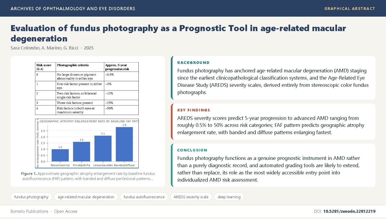
Background: Fundus photography has anchored age-related macular degeneration (AMD) staging since the earliest clinicopathological classification systems, and the Age-Related Eye Disease Study (AREDS) severity scales, derived entirely from stereoscopic color fundus photographs, remain the basis for multi-year risk stratification [5,7].
Objective: This review evaluates the evidence that features visible on fundus photographs, including drusen size, pigmentary abnormality, and fundus autofluorescence (FAF) pattern, carry genuine prognostic value for progression to geographic atrophy and conversion to neovascular AMD.
Methods: We synthesized natural-history cohorts, AREDS and AREDS2 ancillary analyses, FAF pattern studies, and deep-learning re-analyses of legacy fundus photograph archives addressing photographic prediction of AMD progression and vision loss.
Findings: AREDS severity scores predict 5-year progression to advanced AMD ranging from roughly 0.5% to 50% across risk categories [6]; FAF pattern predicts geographic atrophy enlargement rate, with banded and diffuse patterns enlarging fastest [12,11]; and convolutional neural networks trained on archived photographs now match or exceed expert grading, in some cases predicting neovascular conversion months before clinical exudation [25,29].
Conclusions: Fundus photography functions as a genuine prognostic instrument in AMD rather than a purely diagnostic record, and automated grading tools are likely to extend, rather than replace, its role as the most widely accessible entry point into individualized AMD risk assessment [27,26].
References
Wong, W.L., Su, X., Li, X., Cheung, C.M., Klein, R., Cheng, C.Y., et al. (2014) Global prevalence of age-related macular degeneration and disease burden projection for 2020 and 2040: a systematic review and meta-analysis. Lancet Global Health, 2(2): e106-e116. DOI: 10.1016/S2214-109X(13)70145-1
Mitchell, P., Liew, G., Gopinath, B., Wong, T.Y. (2018) Age-related macular degeneration. Lancet, 392(10153): 1147-1159. DOI: 10.1016/S0140-6736(18)31550-2
Lim, L.S., Mitchell, P., Seddon, J.M., Holz, F.G., Wong, T.Y. (2012) Age-related macular degeneration. Lancet, 379(9827): 1728-1738. DOI: 10.1016/S0140-6736(12)60282-7
Bird, A.C., Bressler, N.M., Bressler, S.B., Chisholm, I.H., Coscas, G., Davis, M.D., et al. (1995) An international classification and grading system for age-related maculopathy and age-related macular degeneration. Survey of Ophthalmology, 39(5): 367-374. DOI: 10.1016/S0039-6257(05)80092-X
Age-Related Eye Disease Study Research Group (2001) The Age-Related Eye Disease Study system for classifying age-related macular degeneration from stereoscopic color fundus photographs: AREDS Report Number 6. American Journal of Ophthalmology, 132(5): 668-681. DOI: 10.1016/S0002-9394(01)01218-1
Davis, M.D., Gangnon, R.E., Lee, L.Y., Hubbard, L.D., Klein, B.E., Klein, R., et al. (2005) The Age-Related Eye Disease Study severity scale for age-related macular degeneration: AREDS Report No. 17. Archives of Ophthalmology, 123(11): 1484-1498. DOI: 10.1001/archopht.123.11.1484
Ferris, F.L., Davis, M.D., Clemons, T.E., Lee, L.Y., Chew, E.Y., Lindblad, A.S., et al. (2005) A simplified severity scale for age-related macular degeneration: AREDS Report No. 18. Archives of Ophthalmology, 123(11): 1570-1574. DOI: 10.1001/archopht.123.11.1570
Ferris, F.L., Wilkinson, C.P., Bird, A., Chakravarthy, U., Chew, E., Csaky, K., et al. (2013) Clinical classification of age-related macular degeneration. Ophthalmology, 120(4): 844-851. DOI: 10.1016/j.ophtha.2012.10.036
Klein, R., Klein, B.E., Knudtson, M.D., Meuer, S.M., Swift, M., Gangnon, R.E. (2004) Fifteen-year cumulative incidence of age-related macular degeneration: the Beaver Dam Eye Study. Ophthalmology, 114(2): 253-262.
Klein, R., Meuer, S.M., Knudtson, M.D., Iyengar, S.K., Klein, B.E. (2011) The epidemiology of retinal reticular drusen. American Journal of Ophthalmology, 145(2): 317-326.
Bindewald, A., Bird, A.C., Jung, J.J., Schmitz-Valckenberg, S., Holz, F.G. (2005) Classification of fundus autofluorescence patterns in early age-related macular degeneration. Investigative Ophthalmology & Visual Science, 46(9): 3309-3314. DOI: 10.1167/iovs.04-0430
Holz, F.G., Bindewald-Wittich, A., Fleckenstein, M., Dreyhaupt, J., Scholl, H.P., Schmitz-Valckenberg, S. (2007) Progression of geographic atrophy and impact of fundus autofluorescence patterns in age-related macular degeneration. American Journal of Ophthalmology, 143(3): 463-472. DOI: 10.1016/j.ajo.2006.11.041
Schmitz-Valckenberg, S., Holz, F.G., Bird, A.C., Spaide, R.F. (2008) Fundus autofluorescence imaging: review and perspectives. Retina, 28(3): 385-409. DOI: 10.1097/IAE.0b013e318164a907
Sunness, J.S., Gonzalez-Baron, J., Applegate, C.A., Bressler, N.M., Tian, Y., Hawkins, B., et al. (1999) Enlargement of atrophy and visual acuity loss in the geographic atrophy form of age-related macular degeneration. Ophthalmology, 106(9): 1768-1779. DOI: 10.1016/S0161-6420(99)90397-5
Sunness, J.S., Margalit, E., Srikumaran, D., Applegate, C.A., Tian, Y., Perry, D., et al. (2007) The long-term natural history of geographic atrophy from age-related macular degeneration. Ophthalmology, 114(2): 271-277. DOI: 10.1016/j.ophtha.2006.09.016
Fleckenstein, M., Schmitz-Valckenberg, S., Adrion, C., Bindewald-Wittich, A., Meiβner, S., Bird, A.C., et al. (2011) Concordance of disease progression in bilateral geographic atrophy due to AMD. Investigative Ophthalmology & Visual Science, 51(1): 637-642. DOI: 10.1167/iovs.09-3806
Cukras, C., Agron, E., Klein, M.L., Ferris, F.L., Chew, E.Y., Gensler, G., et al. (2010) Natural history of drusenoid pigment epithelial detachment in age-related macular degeneration: AREDS Report No. 28. Ophthalmology, 117(3): 489-499. DOI: 10.1016/j.ophtha.2009.12.002
Ying, G.S., Maguire, M.G., Daniel, E., Grunwald, J.E., Ahmed, O.M., Klein, M.L., et al. (2013) Baseline predictors for one-year visual acuity outcomes with ranibizumab in patients with neovascular AMD. Ophthalmology, 120(1): 122-129. DOI: 10.1016/j.ophtha.2012.08.001
Rosenfeld, P.J., Brown, D.M., Heier, J.S., Boyer, D.S., Kaiser, P.K., Chung, C.Y., et al. (2006) Ranibizumab for neovascular age-related macular degeneration. New England Journal of Medicine, 355(14): 1419-1431. DOI: 10.1056/NEJMoa054481
Brown, D.M., Kaiser, P.K., Michels, M., Soubrane, G., Heier, J.S., Kim, R.Y., et al. (2006) Ranibizumab versus verteporfin for neovascular age-related macular degeneration. New England Journal of Medicine, 355(14): 1432-1444. DOI: 10.1056/NEJMoa062655
Keane, P.A., Patel, P.J., Liakopoulos, S., Heussen, F.M., Sadda, S.R., Tufail, A. (2012) Evaluation of age-related macular degeneration with optical coherence tomography. Survey of Ophthalmology, 57(5): 389-414. DOI: 10.1016/j.survophthal.2012.01.006
Munk, M.R., Kashani, A.H., Tadayoni, R., Korobelnik, J.F., Wolf, S., Terheyden, J.H., et al. (2021) Standardization of OCT angiography nomenclature in retinal vascular diseases: first survey results. Ophthalmology Retina, 5(10): 981-990.
Seddon, J.M., Rosner, B. (2019) Validated prediction models for macular degeneration progression and predictors of visual acuity loss identify high-risk individuals. American Journal of Ophthalmology, 198: 223-261. DOI: 10.1016/j.ajo.2018.10.022
Burlina, P.M., Joshi, N., Pekala, M., Pacheco, K.D., Freund, D.E., Bressler, N.M. (2017) Automated grading of age-related macular degeneration from color fundus images using deep convolutional neural networks. JAMA Ophthalmology, 135(11): 1170-1176. DOI: 10.1001/jamaophthalmol.2017.3782
Burlina, P.M., Joshi, N., Pacheco, K.D., Freund, D.E., Kong, J., Bressler, N.M. (2018) Use of deep learning for detailed severity characterization and estimation of 5-year risk among patients with age-related macular degeneration. JAMA Ophthalmology, 136(12): 1359-1366. DOI: 10.1001/jamaophthalmol.2018.4118
Grassmann, F., Mengelkamp, J., Brandl, C., Harsch, S., Zimmermann, M.E., Linkohr, B., et al. (2018) A deep learning algorithm for prediction of age-related eye disease study severity scale for age-related macular degeneration from color fundus photography. Ophthalmology, 125(9): 1410-1420. DOI: 10.1016/j.ophtha.2018.02.037
Peng, Y., Dharssi, S., Chen, Q., Keenan, T.D., Agron, E., Wong, W.T., et al. (2019) DeepSeeNet: a deep learning model for automated classification of patient-based age-related macular degeneration severity from color fundus photographs. Ophthalmology, 126(4): 565-575. DOI: 10.1016/j.ophtha.2018.11.015
Schmidt-Erfurth, U., Waldstein, S.M., Klimscha, S., Sadeghipour, A., Hu, X., Gerendas, B.S., et al. (2018) Prediction of individual disease conversion in early AMD using artificial intelligence. Investigative Ophthalmology & Visual Science, 59(8): 3199-3208. DOI: 10.1167/iovs.18-24106
Yim, J., Chopra, R., Spitz, T., Winkens, J., Obika, A., Kelly, C., et al. (2020) Predicting conversion to wet age-related macular degeneration using deep learning. Nature Medicine, 26(6): 892-899. DOI: 10.1038/s41591-020-0867-7
Keenan, T.D., Dharssi, S., Peng, Y., Chen, Q., Agron, E., Wong, W.T., et al. (2020) A deep learning approach for automated detection of geographic atrophy from color fundus photographs. Ophthalmology, 127(11): 1533-1543. DOI: 10.1016/j.ophtha.2020.04.037
Ting, D.S.W., Peng, L., Varadarajan, A.V., Keane, P.A., Burlina, P.M., Chiang, M.F., et al. (2019) Deep learning in ophthalmology: the technical and clinical considerations. Progress in Retinal and Eye Research, 72: 100759. DOI: 10.1016/j.preteyeres.2019.04.003
Age-Related Eye Disease Study Research Group (2001) A randomized, placebo-controlled, clinical trial of high-dose supplementation with vitamins C and E, beta carotene, and zinc for age-related macular degeneration and vision loss: AREDS Report No. 8. Archives of Ophthalmology, 119(10): 1417-1436. DOI: 10.1001/archopht.119.10.1417
Chew, E.Y., Clemons, T.E., Agron, E., Sperduto, R.D., Sangiovanni, J.P., Kurinij, N., et al. (2013) Long-term effects of vitamins C and E, beta-carotene, and zinc on age-related macular degeneration: AREDS Report No. 35. Ophthalmology, 120(8): 1604-1611. DOI: 10.1016/j.ophtha.2013.01.021
Age-Related Eye Disease Study 2 Research Group (2013) Lutein + zeaxanthin and omega-3 fatty acids for age-related macular degeneration: the Age-Related Eye Disease Study 2 randomized clinical trial. JAMA, 309(19): 2005-2015. DOI: 10.1001/jama.2013.4997
Age-Related Eye Disease Study 2 Research Group (2012) The Age-Related Eye Disease Study 2 (AREDS2): study design and baseline characteristics. Ophthalmology, 119(11): 2282-2289. DOI: 10.1016/j.ophtha.2012.05.027
Age-Related Eye Disease Study Research Group (2000) Risk factors associated with age-related macular degeneration: AREDS Report No. 3. Ophthalmology, 107(12): 2224-2232. DOI: 10.1016/S0161-6420(00)00409-7
Age-Related Eye Disease Study Research Group (2005) The relationship of dietary carotenoid and vitamin A, E, and C intake with age-related macular degeneration in a case-control study: AREDS Report No. 22. Archives of Ophthalmology, 125(9): 1225-1232.
Ferris, F.L., Bressler, N.M. (1997) Age-related macular degeneration and clinical trials involving antioxidants and zinc. Archives of Ophthalmology, 115(9): 1222-1223.

Published
Issue
Section
License
Copyright (c) 2025 Sara Colombo, et al.

This work is licensed under a Creative Commons Attribution 4.0 International License.