
Emerging Role of optical coherence tomography in the Diagnosis of diabetic retinopathy
DOI:
https://doi.org/10.5281/zenodo.22812176Keywords:
optical coherence tomography, OCT angiography, diabetic retinopathy, diabetic macular edema, deep learningAbstract
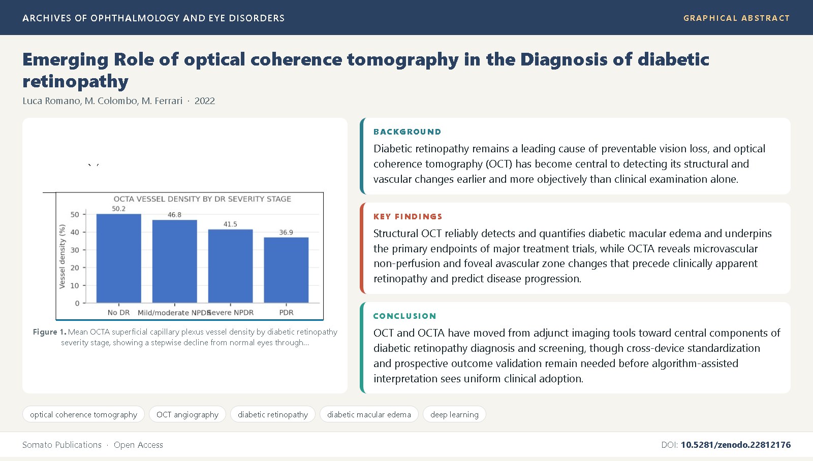
Background: Diabetic retinopathy remains a leading cause of preventable vision loss, and optical coherence tomography (OCT) has become central to detecting its structural and vascular changes earlier and more objectively than clinical examination alone [2,3].
Objective: This review synthesizes the published evidence on structural OCT and OCT angiography (OCTA) in the diagnosis, staging, and monitoring of diabetic retinopathy and diabetic macular edema.
Methods: We synthesized landmark OCT validation studies, randomized treatment trials using OCT-based endpoints, quantitative OCTA cohort studies, and papers on automated and deep-learning-assisted OCT/OCTA interpretation.
Findings: Structural OCT reliably detects and quantifies diabetic macular edema and underpins the primary endpoints of major treatment trials [6,8,9], while OCTA reveals microvascular non-perfusion and foveal avascular zone changes that precede clinically apparent retinopathy and predict disease progression [23,25]; deep-learning systems applied to OCT and OCTA are increasingly able to detect referable disease at accuracy approaching human graders [28,32].
Conclusions: OCT and OCTA have moved from adjunct imaging tools toward central components of diabetic retinopathy diagnosis and screening, though cross-device standardization and prospective outcome validation remain needed before algorithm-assisted interpretation sees uniform clinical adoption [13,30].
References
Yau, J.W.Y., Rogers, S.L., Kawasaki, R., Lamoureux, E.L., Kowalski, J.W., Bek, T., et al. (2012) Global prevalence and major risk factors of diabetic retinopathy. Diabetes Care, 35(3): 556-564. DOI: 10.2337/dc11-1909
Wong, T.Y., Cheung, C.M.G., Larsen, M., Sharma, S., Simo, R. (2016) Diabetic retinopathy. Nature Reviews Disease Primers, 2: 16012. DOI: 10.1038/nrdp.2016.12
Cheung, N., Mitchell, P., Wong, T.Y. (2010) Diabetic retinopathy. Lancet, 376(9735): 124-136. DOI: 10.1016/S0140-6736(09)62124-3
Early Treatment Diabetic Retinopathy Study Research Group (1991) Grading diabetic retinopathy from stereoscopic color fundus photographs--an extension of the modified Airlie House classification. ETDRS report number 10. Ophthalmology, 98(5 Suppl): 786-806. DOI: 10.1016/S0161-6420(13)38012-9
Huang, D., Swanson, E.A., Lin, C.P., Schuman, J.S., Stinson, W.G., Chang, W., et al. (1991) Optical coherence tomography. Science, 254(5035): 1178-1181. DOI: 10.1126/science.1957169
Hee, M.R., Puliafito, C.A., Duker, J.S., Reichel, E., Coker, J.G., Wilkins, J.R., et al. (1998) Topography of diabetic macular edema with optical coherence tomography. Ophthalmology, 105(2): 360-370. DOI: 10.1016/S0161-6420(98)93601-6
Browning, D.J., McOwen, M.D., Bowen, R.M., O'Marah, T.L. (2004) Comparison of the clinical diagnosis of diabetic macular edema with diagnosis by optical coherence tomography. Ophthalmology, 111(4): 712-715. DOI: 10.1016/j.ophtha.2003.06.028
Diabetic Retinopathy Clinical Research Network (2007) Optical coherence tomography and fundus photographic results of a randomized clinical trial comparing intravitreal triamcinolone acetonide and focal/grid photocoagulation for diabetic macular edema. Ophthalmology, 114(9): 1698-1707. DOI: 10.1016/j.ophtha.2007.06.032
Nguyen, Q.D., Brown, D.M., Marcus, D.M., Boyer, D.S., Patel, S., Feiner, L., et al. (2012) Ranibizumab for diabetic macular edema: results from 2 phase III randomized trials, RISE and RIDE. Ophthalmology, 119(4): 789-801. DOI:
1016/j.ophtha.2011.12.039 10. Elman, M.J., Aiello, L.P., Beck, R.W., Bressler, N.M., Bressler, S.B., Edwards, A.R., et al. (2010) Randomized trial evaluating ranibizumab plus prompt or deferred laser or triamcinolone plus prompt laser for diabetic macular edema. Ophthalmology, 117(6): 1064-1077. DOI: 10.1016/j.ophtha.2010.02.031
Wells, J.A., Glassman, A.R., Ayala, A.R., Jampol, L.M., Bressler, N.M., Bressler, S.B., et al. (2016) Aflibercept, bevacizumab, or ranibizumab for diabetic macular edema: two-year results from a comparative effectiveness randomized clinical trial. Ophthalmology, 123(6): 1351-1359. DOI: 10.1016/j.ophtha.2016.02.022
Sadda, S.R., Wu, Z., Walsh, A.C., Richine, L., Dougall, J., Cortez, R., et al. (2006) Errors in retinal thickness measurements obtained by optical coherence tomography. Ophthalmology, 113(2): 285-293. DOI: 10.1016/j.ophtha.2005.10.005
Virgili, G., Menchini, F., Casazza, G., Hogg, R., Das, R.R., Wang, X., et al. (2015) Optical coherence tomography (OCT) for detection of macular oedema in patients with diabetic retinopathy. Cochrane Database of Systematic Reviews, (1): CD008081. DOI: 10.1002/14651858.CD008081.pub3
Gross, J.G., Glassman, A.R., Jampol, L.M., Inusah, S., Aiello, L.P., Antoszyk, A.N., et al. (2015) Panretinal photocoagulation vs intravitreous ranibizumab for proliferative diabetic retinopathy. JAMA, 314(20): 2137-2146. DOI: 10.1001/jama.2015.15217
Spaide, R.F., Fujimoto, J.G., Waheed, N.K., Sadda, S.R., Staurenghi, G. (2018) Optical coherence tomography angiography. Progress in Retinal and Eye Research, 64: 1-55. DOI: 10.1016/j.preteyeres.2017.11.003
Spaide, R.F., Fujimoto, J.G., Waheed, N.K. (2015) Image artifacts in optical coherence tomography angiography. Retina, 35(11): 2163-2180. DOI: 10.1097/IAE.0000000000000765
Ishibazawa, A., Nagaoka, T., Takahashi, A., Omae, T., Tani, T., Sogawa, K., et al. (2015) Optical coherence tomography angiography in diabetic retinopathy: a prospective pilot study. American Journal of Ophthalmology, 160(1): 35-44. DOI: 10.1016/j.ajo.2015.04.021
Soares, M., Neves, C., Marques, I.P., Pires, I., Schwartz, C., Costa, M.A., et al. (2017) Comparison of diabetic retinopathy classification using fluorescein angiography and optical coherence tomography angiography. British Journal of Ophthalmology, 101(1): 62-68. DOI: 10.1136/bjophthalmol-2016-309424
Ting, D.S.W., Tan, G.S.W., Agrawal, R., Yanagi, Y., Sie, N.M., Wong, C.W., et al. (2015) Optical coherence tomographic angiography in type 2 diabetes and diabetic retinopathy. JAMA Ophthalmology, 133(8): 986-993. DOI: 10.1001/jamaophthalmol.2015.1970
Agemy, S.A., Scripsema, N.K., Shah, C.M., Chui, T., Garcia, P.M., Lee, J.G., et al. (2015) Retinal vascular perfusion density mapping using optical coherence tomography angiography in normals and diabetic retinopathy patients. Retina, 35(11): 2353-2363. DOI: 10.1097/IAE.0000000000000862
Kim, A.Y., Chu, Z., Shahidzadeh, A., Wang, R.K., Puliafito, C.A., Kashani, A.H. (2016) Quantifying microvascular density and morphology in diabetic retinopathy using spectral-domain optical coherence tomography angiography. Investigative Ophthalmology & Visual Science, 57(9): OCT362-OCT370. DOI: 10.1167/iovs.15-18904
Durbin, M.K., An, L., Shemonski, N.D., Soares, M., Santos, T., Lopes, M., et al. (2017) Quantification of retinal microvascular density in optical coherence tomographic angiography images in diabetic retinopathy. JAMA Ophthalmology, 135(4): 370-376. DOI: 10.1001/jamaophthalmol.2017.0080
Cao, D., Yang, D., Yang, D., Cheng, C., Zhang, L., Yang, Z., et al. (2018) Optical coherence tomography angiography discerns preclinical diabetic retinopathy in eyes of patients with type 2 diabetes without clinical diabetic retinopathy. Acta Diabetologica, 55(5): 469-477. DOI: 10.1007/s00592-018-1115-1
de Carlo, T.E., Chin, A.T., Bonini Filho, M.A., Adhi, M., Branchini, L., Salz, D.A., et al. (2015) Detection of microvascular changes in eyes of patients with diabetes but not clinical diabetic retinopathy using optical coherence tomography angiography. Retina, 35(11): 2364-2370. DOI: 10.1097/IAE.0000000000000882
Sun, Z., Tang, F., Wong, R., Lok, J., Szeto, S.K.H., Chan, J.C.K., et al. (2019) OCT angiography metrics predict progression of diabetic retinopathy and development of diabetic macular edema. Ophthalmology, 126(12): 1675-1684. DOI: 10.1016/j.ophtha.2019.06.016
Sun, Z., Yang, D., Tang, Z., Ng, D.S., Cheung, C.Y. (2021) Optical coherence tomography angiography in diabetic retinopathy: an updated review. Eye, 35(1): 149-161. DOI: 10.1038/s41433-020-01233-y
Gill, A., Cole, E.D., Novais, E.A., Louzada, R.N., Waheed, N.K., Regatieri, C.V., et al. (2017) Visualization of changes in the foveal avascular zone in both observed and treated diabetic macular edema using optical coherence tomography angiography. International Journal of Retina and Vitreous, 3: 19. DOI: 10.1186/s40942-017-0068-9
Gulshan, V., Peng, L., Coram, M., Stumpe, M.C., Wu, D., Narayanaswamy, A., et al. (2016) Development and validation of a deep learning algorithm for detection of diabetic retinopathy in retinal fundus photographs. JAMA, 316(22): 2402-2410. DOI: 10.1001/jama.2016.17216
Ting, D.S.W., Cheung, C.Y., Lim, G., Tan, G.S.W., Quang, N.D., Gan, A., et al. (2017) Development and validation of a deep learning system for diabetic retinopathy and related eye diseases using retinal images from multiethnic populations with diabetes. JAMA, 318(22): 2211-2223. DOI: 10.1001/jama.2017.18152
Heisler, M., Karst, S., Lo, J., Mammo, Z., Yu, T., Warner, S., et al. (2020) Ensemble deep learning for diabetic retinopathy detection using optical coherence tomography angiography. Translational Vision Science & Technology, 9(2): 20. DOI: 10.1167/tvst.9.2.20
Le, D., Alam, M., Yao, C.K., Lim, J.I., Hsieh, Y.T., Chan, R.V.P., et al. (2020) Transfer learning for automated OCTA detection of diabetic retinopathy. Translational Vision Science & Technology, 9(2): 35. DOI: 10.1167/tvst.9.2.35
Zhang, Y., Fu, D., Liu, T., Chen, M., Zheng, Z., Chen, Y., et al. (2021) Development of an automated diabetic macular edema detection system using optical coherence tomography images and deep learning. Journal of Diabetes Research, 2021: 1394832. DOI: 10.1155/2021/1394832

Published
Issue
Section
License
Copyright (c) 2022 Luca Romano, et al.

This work is licensed under a Creative Commons Attribution 4.0 International License.