Predictors of cellular uptake efficiency in preclinical animal models with bacterial infections
DOI:
https://doi.org/10.5281/zenodo.22811896Keywords:
cellular uptake, nanoparticle, bacterial infection, protein corona, biofilmAbstract
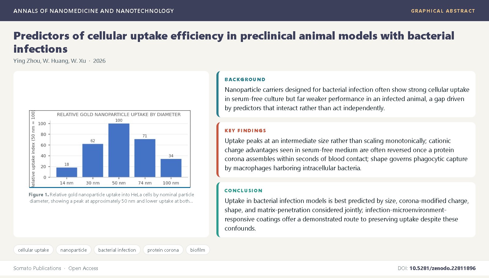
Background: Nanoparticle carriers designed for bacterial infection often show strong cellular uptake in serum-free culture but far weaker performance in an infected animal, a gap driven by predictors that interact rather than act independently.
Objective: This review synthesizes evidence on what predicts nanoparticle cellular uptake efficiency in preclinical bacterial infection models, spanning size, charge, shape, protein corona formation, and the biofilm barrier unique to infection.
Methods: We conducted a narrative synthesis of foundational biophysical uptake studies and infection-specific studies of nanoparticle transport in murine and biofilm models.
Findings: Uptake peaks at an intermediate size rather than scaling monotonically [4]; cationic charge advantages seen in serum-free medium are often reversed once a protein corona assembles within seconds of blood contact [9,12]; shape governs phagocytic capture by macrophages harboring intracellular bacteria [13]; and biofilm matrix acts as a size- and charge-selective barrier absent from single-cell assays [19,20].
Conclusions: Uptake in bacterial infection models is best predicted by size, corona-modified charge, shape, and matrix-penetration considered jointly; infection-microenvironment-responsive coatings offer a demonstrated route to preserving uptake despite these confounds [21,22].
References
Wilhelm, S., Tavares, A.J., Dai, Q., Ohta, S., Audet, J., Dvorak, H.F., et al. (2016) Analysis of nanoparticle delivery to tumours. Nature Reviews Materials, 1: 16014. DOI: 10.1038/natrevmats.2016.14
Nel, A.E., Madler, L., Velegol, D., Xia, T., Hoek, E.M.V., Somasundaran, P., et al. (2009) Understanding biophysicochemical interactions at the nano-bio interface. Nature Materials, 8(7): 543-557. DOI: 10.1038/nmat2442
Albanese, A., Tang, P.S., Chan, W.C.W. (2012) The effect of nanoparticle size, shape, and surface chemistry on biological systems. Annual Review of Biomedical Engineering, 14: 1-16. DOI: 10.1146/annurev-bioeng-071811-150124
Chithrani, B.D., Ghazani, A.A., Chan, W.C.W. (2006) Determining the size and shape dependence of gold nanoparticle uptake into mammalian cells. Nano Letters, 6(4): 662-668. DOI: 10.1021/nl0523960
Chithrani, B.D., Chan, W.C.W. (2007) Elucidating the mechanism of cellular uptake and removal of protein-coated gold nanoparticles of different sizes and shapes. Nano Letters, 7(6): 1542-1550. DOI: 10.1021/nl070363y
Jiang, W., Kim, B.Y.S., Rutka, J.T., Chan, W.C.W. (2008) Nanoparticle-mediated cellular response is size-dependent. Nature Nanotechnology, 3(3): 145-150. DOI: 10.1038/nnano.2008.30
Frohlich, E. (2012) The role of surface charge in cellular uptake and cytotoxicity of medical nanoparticles. International Journal of Nanomedicine, 7: 5577-5591. DOI: 10.2147/IJN.S36111
Verma, A., Stellacci, F. (2010) Effect of surface properties on nanoparticle-cell interactions. Small, 6(1): 12-21. DOI: 10.1002/smll.200901158
Walkey, C.D., Olsen, J.B., Guo, H., Emili, A., Chan, W.C.W. (2012) Nanoparticle size and surface chemistry determine serum protein adsorption and macrophage uptake. Journal of the American Chemical Society, 134(4): 2139-2147. DOI:
1021/ja2084338 10. Cedervall, T., Lynch, I., Lindman, S., Berggard, T., Thulin, E., Nilsson, H., et al. (2007) Understanding the nanoparticle-protein corona using methods to quantify exchange rates and affinities of proteins. Proceedings of the National Academy of Sciences, 104(7): 2050-2055. DOI: 10.1073/pnas.0608582104
Monopoli, M.P., Aberg, C., Salvati, A., Dawson, K.A. (2012) Biomolecular coronas provide the biological identity of nanosized materials. Nature Nanotechnology, 7(12): 779-786. DOI: 10.1038/nnano.2012.207
Tenzer, S., Docter, D., Kuharev, J., Musyanovych, A., Fetz, V., Hecht, R., et al. (2013) Rapid formation of plasma protein corona critically affects nanoparticle pathophysiology. Nature Nanotechnology, 8(10): 772-781. DOI: 10.1038/nnano.2013.181
Champion, J.A., Mitragotri, S. (2006) Role of target geometry in phagocytosis. Proceedings of the National Academy of Sciences, 103(13): 4930-4934. DOI: 10.1073/pnas.0600997103
Gustafson, H.H., Holt-Casper, D., Grainger, D.W., Ghandehari, H. (2015) Nanoparticle uptake: the phagocyte problem. Nano Today, 10(4): 487-510. DOI: 10.1016/j.nantod.2015.06.006
Kettler, K., Veltman, K., van de Meent, D., van Wezel, A., Hendriks, A.J. (2014) Cellular uptake of nanoparticles as determined by particle size, surface shape, and cell type. Environmental Toxicology and Chemistry, 33(3): 481-492. DOI: 10.1002/etc.2470
Thorley, A.J., Ruenraroengsak, P., Potter, T.J., Tetley, T.D. (2014) Critical determinants of uptake and translocation of nanoparticles by the human pulmonary alveolar epithelium. ACS Nano, 8(11): 11778-11789. DOI: 10.1021/nn505399e
Foroozandeh, P., Aziz, A.A. (2018) Insight into cellular uptake and intracellular trafficking of nanoparticles. Nanoscale Research Letters, 13: 339. DOI: 10.1186/s11671-018-2728-6
Behzadi, S., Serpooshan, V., Tao, W., Hamaly, M.A., Alkawareek, M.Y., Dreaden, E.C., et al. (2017) Cellular uptake of nanoparticles: journey inside the cell. Chemical Society Reviews, 46(14): 4218-4244. DOI: 10.1039/c6cs00636a
Forier, K., Raemdonck, K., De Smedt, S.C., Demeester, J., Coenye, T., Braeckmans, K. (2014) Transport of nanoparticles in cystic fibrosis sputum and bacterial biofilms by single-particle tracking microscopy. Nanomedicine, 9(15): 2417-2432. DOI: 10.2217/nnm.14.155
Baelo, A., Levato, R., Julian, E., Crespo, A., Astola, J., Gavalda, J., et al. (2015) Disassembling bacterial extracellular matrix with DNase-coated nanoparticles to enhance antibiotic delivery in biofilm infections. Journal of Controlled Release, 209: 150-158. DOI: 10.1016/j.jconrel.2015.04.028
Radovic-Moreno, A.F., Lu, T.K., Puscasu, V.A., Yoon, C.J., Langer, R., Farokhzad, O.C. (2012) Surface charge-switching polymeric nanoparticles for bacterial cell wall-targeted delivery of antibiotics. ACS Nano, 6(5): 4279-4287. DOI: 10.1021/nn3010918
Xiong, M.H., Bao, Y., Yang, X.Z., Zhu, Y.H., Wang, J. (2012) Lipase-sensitive polymeric triple-layered nanogel for "on-demand" drug delivery. Journal of the American Chemical Society, 134(9): 4355-4362. DOI: 10.1021/ja211279u
Panizzi, P., Nahrendorf, M., Figueiredo, J.L., Panizzi, J., Marinelli, B., Iwamoto, Y., et al. (2011) In vivo detection of Staphylococcus aureus endocarditis by targeting pathogen-specific prothrombin activation. Nature Medicine, 17(9): 1142-1146. DOI: 10.1038/nm.2423
Owens, D.E., Peppas, N.A. (2006) Opsonization, biodistribution, and pharmacokinetics of polymeric nanoparticles. International Journal of Pharmaceutics, 307(1): 93-102. DOI: 10.1016/j.ijpharm.2005.10.010
Blanco, E., Shen, H., Ferrari, M. (2015) Principles of nanoparticle design for overcoming biological barriers to drug delivery. Nature Biotechnology, 33(9): 941-951. DOI: 10.1038/nbt.3330
Iversen, T.G., Skotland, T., Sandvig, K. (2011) Endocytosis and intracellular transport of nanoparticles: present knowledge and need for future studies. Nano Today, 6(2): 176-185. DOI: 10.1016/j.nantod.2011.02.003
Zhao, J., Stenzel, M.H. (2018) Entry of nanoparticles into cells: the importance of nanoparticle properties. Polymer Chemistry, 9(3): 259-272. DOI: 10.1039/C7PY01603D

Published
Issue
Section
License
Copyright (c) 2026 Ying Zhou, et al.

This work is licensed under a Creative Commons Attribution 4.0 International License.